Developing and validating an elderly brain template: A comprehensive comparison with MNI152 for age-specific neuroimaging analyses
IF 4.5
2区 医学
Q1 NEUROIMAGING
引用次数: 0
Abstract
Background
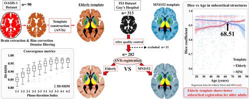
The Montreal Neurological Institute 152 (MNI152) brain template, constructed from young adult brains, may not accurately represent older age–specific morphological changes. Accordingly, we developed and validated the new Elderly template.
Methods
MRI scans from 90 OASIS-1 participants, matching Japanese census demographics, were used to construct the Elderly template. Spatial normalization accuracy was compared with that of the MNI152 in the IXI dataset. Following UK Biobank–based intracranial volume quality control (±2 SD; 1232–1850 mL), 282 of 313 scans from individuals aged 20–80+ years were included. Whole-brain similarity was assessed with cross-correlation (CC), mean-squared error (MSE), and 3D structural similarity index measure (3D-SSIM). Dice coefficients were computed for white matter (WM), gray matter (GM), cerebrospinal fluid (CSF), and seven subcortical regions. Generalized linear models were used to test the Age × Template interactions (β₃).
Results
Significant Age × Template interactions were observed for CC and MSE (p < 0.001); 3D-SSIM showed a positive but non-significant trend. Dice analyses mirrored this pattern: WM and GM showed minor differences between templates, and the Dice coefficient was parallel. CSF showed a sharp difference at the age ≥60 years. The largest interaction effects (≈1–4 % gain) occurred in the caudate, thalamus, hippocampus, and amygdala, whereas the brainstem, pallidum, and putamen showed minimal differences between templates.
Conclusions
The Elderly template more accurately reflects older age–specific morphological changes and enhances spatial normalization accuracy, compared with the MNI152 template. This improvement suggests advancements in age-specific analyses and neurodegenerative disease research, enabling clinical applications.
开发和验证老年人大脑模板:与MNI152进行年龄特异性神经影像学分析的综合比较。
背景:蒙特利尔神经学研究所152 (MNI152)脑模板,构建自年轻成人的大脑,可能不能准确地代表年龄特异性的形态学变化。因此,我们开发并验证了新的老年人模板。方法:使用符合日本人口普查统计数据的90名OASIS-1参与者的MRI扫描来构建老年人模板。将空间归一化精度与IXI数据集中的MNI152进行比较。根据基于UK biobank的颅内体积质量控制(±2 SD; 1232-1850 mL),来自20-80岁以上个体的313次扫描中有282次被纳入。采用互相关(CC)、均方误差(MSE)和3D结构相似性指数测量(3D- ssim)评估全脑相似性。计算白质(WM)、灰质(GM)、脑脊液(CSF)和7个皮质下区域的骰子系数。采用广义线性模型对Age × 模板相互作用(β₃)进行检验。结果:CC和MSE存在显著的年龄 × 模板相互作用(p < 0.001);3D-SSIM呈阳性但不显著趋势。骰子分析反映了这一模式:WM和GM显示出模板之间的微小差异,骰子系数是平行的。CSF在≥60岁时表现出明显的差异。最大的相互作用效应(≈1-4%的增益)发生在尾状体、丘脑、海马和杏仁核,而脑干、白质和壳核在模板之间的差异很小。结论:与MNI152模板相比,老年模板更准确地反映了老年人特异性形态学变化,并提高了空间归一化精度。这一改进表明了年龄特异性分析和神经退行性疾病研究的进步,使临床应用成为可能。
本文章由计算机程序翻译,如有差异,请以英文原文为准。
求助全文
约1分钟内获得全文
求助全文
来源期刊

NeuroImage
医学-核医学
CiteScore
11.30
自引率
10.50%
发文量
809
审稿时长
63 days
期刊介绍:
NeuroImage, a Journal of Brain Function provides a vehicle for communicating important advances in acquiring, analyzing, and modelling neuroimaging data and in applying these techniques to the study of structure-function and brain-behavior relationships. Though the emphasis is on the macroscopic level of human brain organization, meso-and microscopic neuroimaging across all species will be considered if informative for understanding the aforementioned relationships.
 求助内容:
求助内容: 应助结果提醒方式:


