Impact of electrode–tissue proximity on formation of pulsed field ablation lesions: Using multi-electrode impedance measurements
IF 2.9
Q2 CARDIAC & CARDIOVASCULAR SYSTEMS
引用次数: 0
Abstract
Background
For pulsed field ablation (PFA), the effect of electrode–tissue contact or proximity remains unclear.
Objective
In this porcine study, the effect of electrode–tissue proximity was studied after 3 weeks of follow-up, using a custom device in which the electrode–tissue distance could be varied for every electrode.
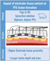
Methods
An impedance-based method was used to determine electrode–tissue proximity or contact. During a bench experiment, the relation between contact value per electrode and electrode–tissue distance was studied. In addition, in 10 pigs epicardial ablation using a custom 6-polar linear ablation device was performed using bipolar, biphasic deliveries on randomized locations on the heart. After 3 weeks, macroscopic analysis was used to compare lesion width for left vs right ventricle. Based on histology, the lesion area and depth were related to electrode–tissue contact.
Results
Analysis of 96 bench measurements showed a decrease in tissue-proximity index with increasing distance. Lesions on the right ventricle were wider compared to the lesions on the left ventricle, 9.0 ± 1.9 mm vs 6.5 ± 1.0 mm (P < .001), respectively. A total of 162 lesions showed that a higher tissue-proximity index was associated with deeper and larger lesions; lesion depth was 1.3 ± 0.5 mm vs 1.5 ± 0.5 mm (P = .016) and lesion area 6.4 ± 2.7 mm2 vs 8.3 ± 2.9 mm2 (P < .001) for low vs high indices respectively.
Conclusion
This study shows that higher electrode–tissue proximity may result in deeper PFA lesions, and is, therefore, an important parameter in improving PFA lesions. Therefore, confirmation of tissue contact using impedance-based measurements may improve PFA ablation efficacy in a clinical setting.
电极-组织接近对脉冲场消融病变形成的影响:使用多电极阻抗测量
背景:对于脉冲场消融(PFA),电极与组织接触或接近的影响尚不清楚。目的在猪实验中,采用可改变电极与组织距离的定制装置,在随访3周后研究电极与组织距离的影响。方法采用阻抗法测定电极与组织的接近度或接触度。在台架实验中,研究了每个电极的接触值与电极-组织距离的关系。此外,10头猪心外膜消融采用定制的6极线性消融装置,在心脏的随机位置采用双相双相输送。3周后,采用宏观分析比较左、右心室病变宽度。从组织学上看,损伤的面积和深度与电极与组织的接触有关。结果96个台架测量结果显示,组织接近指数随距离的增加而降低。右心室病变较左心室宽,分别为9.0±1.9 mm和6.5±1.0 mm (P < .001)。162个病变表明,较高的组织邻近指数与更深、更大的病变相关;病变深度分别为1.3±0.5 mm vs 1.5±0.5 mm (P = 0.016),病变面积分别为6.4±2.7 mm2 vs 8.3±2.9 mm2 (P < .001)。结论电极组织接近度越高,PFA病变越深,是改善PFA病变的重要参数。因此,使用基于阻抗的测量来确认组织接触可能会提高PFA消融在临床中的疗效。
本文章由计算机程序翻译,如有差异,请以英文原文为准。
求助全文
约1分钟内获得全文
求助全文
来源期刊

Heart Rhythm O2
Cardiology and Cardiovascular Medicine
CiteScore
3.30
自引率
0.00%
发文量
0
审稿时长
52 days
 求助内容:
求助内容: 应助结果提醒方式:


