Intramuscular variations in the exit of C5 and C6 roots of the brachial plexus: A case series from Rwanda
Q3 Medicine
引用次数: 0
Abstract
Background
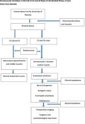
Anatomical variations in the brachial plexus, particularly the relationship of its roots to the scalene muscles, can significantly impact surgical approaches, nerve blocks, and trauma management. However, such variations are underreported in human cadaveric studies from the East African region in general, and Rwanda in particular.
Objective
This case series documents four distinct variations of the brachial plexus identified during routine cadaveric dissection.
Materials and methods
Forty eight brachial plexus (24 human donors) were dissected using Grant's dissector protocol. All specimens were formalin-fixed and dissected in the anatomy laboratory of the University of Rwanda, with ethical clearance obtained.
Results
Four cases (4/48 = 8.33 %) of intramuscular variation in the exit of C5 and C6 were identified. In case 1, right C5 and C6 roots pierced the anterior scalene muscle instead of passing in the scalenic space. Case 2 showed left C5 piercing the anterior scalene. In case 3 and 4 (bilateral variations in one cadaver), C5 and C6 pierced the anterior scalene on both sides but at different locations. These deviations from the classical interscalene passage may pose risks during surgical or anesthetic procedures.
Conclusion
Anatomical variations of the brachial plexus are not rare and warrant routine consideration during clinical procedures. Preoperative imaging and surgeon awareness are recommended to mitigate iatrogenic risks.
臂丛C5和C6根出口的肌内变异:来自卢旺达的一系列病例
臂丛神经的解剖学变异,特别是其根与斜角肌的关系,可以显著影响手术入路、神经阻滞和创伤处理。然而,在整个东非地区,特别是卢旺达的人类尸体研究中,这种差异被低估了。目的:本病例系列记录了在常规尸体解剖中发现的臂丛的四种不同变化。材料和方法采用Grant解剖方案对48例臂丛(24例供体)进行解剖。所有标本都用福尔马林固定,并在卢旺达大学解剖实验室进行解剖,并获得伦理许可。结果发现C5、C6出口肌内变异4例(4/48 = 8.33%)。在病例1中,右侧C5和C6根刺穿前斜角肌而不是穿过斜角肌间隙。病例2显示左侧C5刺穿前斜角肌。病例3和4(同一具尸体的双侧变异),C5和C6刺穿了两侧的前斜角肌,但位置不同。这些与经典斜角肌间通道的偏离可能在手术或麻醉过程中造成危险。结论臂丛神经的解剖变异并不罕见,在临床手术中应予以常规考虑。建议术前影像学检查和外科医生的意识来降低医源性风险。
本文章由计算机程序翻译,如有差异,请以英文原文为准。
求助全文
约1分钟内获得全文
求助全文
来源期刊

Translational Research in Anatomy
Medicine-Anatomy
CiteScore
2.90
自引率
0.00%
发文量
71
审稿时长
25 days
期刊介绍:
Translational Research in Anatomy is an international peer-reviewed and open access journal that publishes high-quality original papers. Focusing on translational research, the journal aims to disseminate the knowledge that is gained in the basic science of anatomy and to apply it to the diagnosis and treatment of human pathology in order to improve individual patient well-being. Topics published in Translational Research in Anatomy include anatomy in all of its aspects, especially those that have application to other scientific disciplines including the health sciences: • gross anatomy • neuroanatomy • histology • immunohistochemistry • comparative anatomy • embryology • molecular biology • microscopic anatomy • forensics • imaging/radiology • medical education Priority will be given to studies that clearly articulate their relevance to the broader aspects of anatomy and how they can impact patient care.Strengthening the ties between morphological research and medicine will foster collaboration between anatomists and physicians. Therefore, Translational Research in Anatomy will serve as a platform for communication and understanding between the disciplines of anatomy and medicine and will aid in the dissemination of anatomical research. The journal accepts the following article types: 1. Review articles 2. Original research papers 3. New state-of-the-art methods of research in the field of anatomy including imaging, dissection methods, medical devices and quantitation 4. Education papers (teaching technologies/methods in medical education in anatomy) 5. Commentaries 6. Letters to the Editor 7. Selected conference papers 8. Case Reports
 求助内容:
求助内容: 应助结果提醒方式:


