Guillaume Fahrni, Salim Si-Mohamed, Rafael Wiemker, David C Rotzinger, Angèle Houmeau, Cyril Prieur, Philippe Douek, Sara Boccalini
{"title":"Spectral photon-counting CT in first-pass myocardial perfusion imaging for very high-risk patients: a comparison with dual-energy CT.","authors":"Guillaume Fahrni, Salim Si-Mohamed, Rafael Wiemker, David C Rotzinger, Angèle Houmeau, Cyril Prieur, Philippe Douek, Sara Boccalini","doi":"10.1186/s41747-025-00624-8","DOIUrl":null,"url":null,"abstract":"<p><strong>Background: </strong>Spectral photon-counting computed tomography (SPCCT) outperformed dual-energy computed tomography (DECT) for coronary artery stenosis assessment. However, data about myocardial perfusion imaging (MPI) is lacking. This feasibility study aimed to evaluate and compare the diagnostic performance of SPCCT and DECT for rest MPI in patients with hemodynamically significant coronary stenoses, using invasive coronary angiography (ICA) and invasive fractional flow reserve (FFR) as reference standards.</p><p><strong>Materials and methods: </strong>Eighteen very-high-risk patients with hemodynamically significant coronary stenoses at ICA underwent both dual-layer DECT and SPCCT coronary CT within three days. The sensitivity, specificity, and accuracy of MPI in detecting myocardial hypoperfusion were assessed. Quantitative attenuation differences between normal and hypoperfused myocardial segments were compared for both modalities. Interobserver variability was assessed with a weighted kappa analysis.</p><p><strong>Results: </strong>SPCCT demonstrated comparable overall performance to DECT for MPI, with an overall sensitivity, specificity, and accuracy of 73.3%, 79.2%, and 76.9%, respectively, versus 73.3%, 75%, and 74.4% for DECT. SPCCT outperformed DECT in the left anterior descending artery territory, achieving a sensitivity of 87.5%, specificity of 100%, and accuracy of 90%, versus 62.5%, 50%, and 60% for DECT. For each CT system, attenuation analysis revealed differences between normal and hypoperfused segments, with mean differences of 17.9 HU for DECT and 15.8 HU for SPCCT (p < 0.05). Inter-reader agreement was higher for SPCCT (κ = 0.86) compared to DECT (κ = 0.62).</p><p><strong>Conclusion: </strong>SPCCT and DECT provided similar diagnostic performance for rest MPI in a very-high-risk patient cohort, demonstrating comparable effectiveness in detecting the effects of hemodynamically significant coronary stenosis.</p><p><strong>Relevance statement: </strong>Hemodynamically significant stenosis in very-high-risk patients results in myocardial hypoperfused areas at rest that can be detected equally well with dual-layer CT and spectral photon counting CT, albeit with better reproducibility for the latter.</p><p><strong>Key points: </strong>SPCCT and DECT showed comparable performance for MPI at rest in very-high-risk patients. The differences between normal and hypoperfused segments were of 17 HU and 16 HU on conventional images for DECT and SPCCT. SPCCT showed higher interobserver agreement compared to DECT, suggesting improved reproducibility.</p>","PeriodicalId":36926,"journal":{"name":"European Radiology Experimental","volume":"9 1","pages":"94"},"PeriodicalIF":3.6000,"publicationDate":"2025-09-20","publicationTypes":"Journal Article","fieldsOfStudy":null,"isOpenAccess":false,"openAccessPdf":"https://www.ncbi.nlm.nih.gov/pmc/articles/PMC12450193/pdf/","citationCount":"0","resultStr":null,"platform":"Semanticscholar","paperid":null,"PeriodicalName":"European Radiology Experimental","FirstCategoryId":"1085","ListUrlMain":"https://doi.org/10.1186/s41747-025-00624-8","RegionNum":0,"RegionCategory":null,"ArticlePicture":[],"TitleCN":null,"AbstractTextCN":null,"PMCID":null,"EPubDate":"","PubModel":"","JCR":"Q1","JCRName":"RADIOLOGY, NUCLEAR MEDICINE & MEDICAL IMAGING","Score":null,"Total":0}
引用次数: 0
Abstract
Background: Spectral photon-counting computed tomography (SPCCT) outperformed dual-energy computed tomography (DECT) for coronary artery stenosis assessment. However, data about myocardial perfusion imaging (MPI) is lacking. This feasibility study aimed to evaluate and compare the diagnostic performance of SPCCT and DECT for rest MPI in patients with hemodynamically significant coronary stenoses, using invasive coronary angiography (ICA) and invasive fractional flow reserve (FFR) as reference standards.
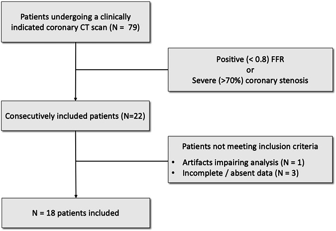
Materials and methods: Eighteen very-high-risk patients with hemodynamically significant coronary stenoses at ICA underwent both dual-layer DECT and SPCCT coronary CT within three days. The sensitivity, specificity, and accuracy of MPI in detecting myocardial hypoperfusion were assessed. Quantitative attenuation differences between normal and hypoperfused myocardial segments were compared for both modalities. Interobserver variability was assessed with a weighted kappa analysis.
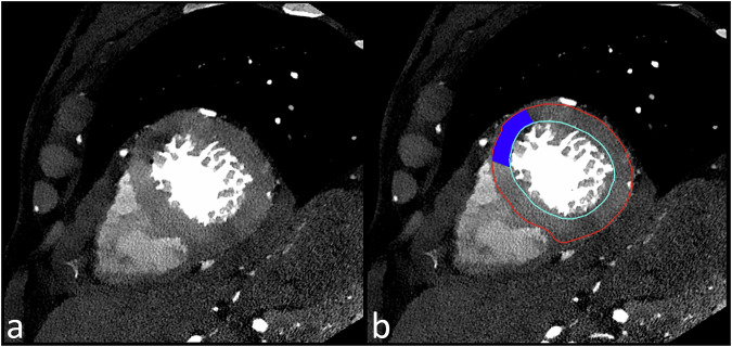
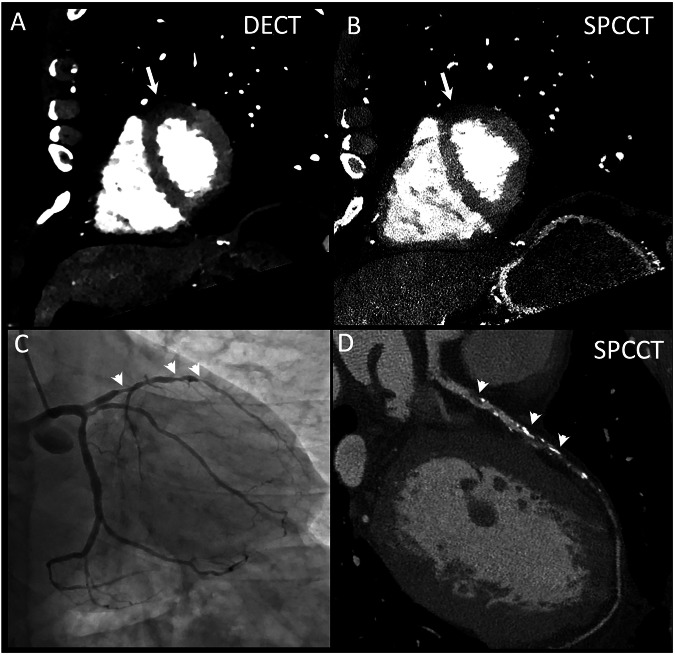
Results: SPCCT demonstrated comparable overall performance to DECT for MPI, with an overall sensitivity, specificity, and accuracy of 73.3%, 79.2%, and 76.9%, respectively, versus 73.3%, 75%, and 74.4% for DECT. SPCCT outperformed DECT in the left anterior descending artery territory, achieving a sensitivity of 87.5%, specificity of 100%, and accuracy of 90%, versus 62.5%, 50%, and 60% for DECT. For each CT system, attenuation analysis revealed differences between normal and hypoperfused segments, with mean differences of 17.9 HU for DECT and 15.8 HU for SPCCT (p < 0.05). Inter-reader agreement was higher for SPCCT (κ = 0.86) compared to DECT (κ = 0.62).
Conclusion: SPCCT and DECT provided similar diagnostic performance for rest MPI in a very-high-risk patient cohort, demonstrating comparable effectiveness in detecting the effects of hemodynamically significant coronary stenosis.
Relevance statement: Hemodynamically significant stenosis in very-high-risk patients results in myocardial hypoperfused areas at rest that can be detected equally well with dual-layer CT and spectral photon counting CT, albeit with better reproducibility for the latter.
Key points: SPCCT and DECT showed comparable performance for MPI at rest in very-high-risk patients. The differences between normal and hypoperfused segments were of 17 HU and 16 HU on conventional images for DECT and SPCCT. SPCCT showed higher interobserver agreement compared to DECT, suggesting improved reproducibility.

 求助内容:
求助内容: 应助结果提醒方式:


