Single cell analysis identifies a distinct population of fibroblasts that mediate increased cell-cell communication in murine aortopathy of Loeys-Dietz syndrome
IF 4.7
2区 医学
Q1 CARDIAC & CARDIOVASCULAR SYSTEMS
引用次数: 0
Abstract
Background
Loeys-Dietz syndrome (LDS), caused by heterozygous loss-of-function mutations in members of transforming growth factor β (TGFβ) pathway, results in frequent aortic root aneurysms and type A dissections in human.
Methods
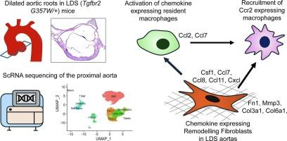
To unveil the mechanism of pathogenesis, the present study utilized single cell RNA sequencing (scRNAseq) from the proximal aortas (aortic root and ascending aorta) of 20 weeks old LDS (Tgfbr2G357W/+) and wild type (Tgfbr2+/+) mice. Histological and immunofluorescence studies were performed on 30 weeks old mice.
Results
ScRNAseq study identifies presence of an exclusive fibroblast population (remodeling fibroblasts) in the proximal aortas of LDS mice, which differentially expressed increased extracellular matrix remodeling genes (Mmp3, Col6a5, Col3a1, and Fn1), and macrophage recruiting chemokines (Saa3, Ccl7, Ccl8, and Cxcl11). These remodeling fibroblasts are focally localized with macrophages at the adventitia of dilated aortic roots of LDS mice. LDS aortas showed increased accumulation of Ccr2 expressing infiltrating macrophages, which are functionally involved in phagocytosis, immune responses and antigen processing and presentation. Ligand-receptor based interaction model recognizes remodeling fibroblasts as a major mediator for signaling communications with resident and recruited macrophages in the proximal aortopathies of LDS mice.
Conclusion
Our study highlights the presence of a specialized fibroblast population in the dilated aortic roots of LDS mice at 20 weeks and provides a deeper insight for involvement of remodeling fibroblasts in cellular heterogeneity and cell-cell communications in LDS aortopathies.
单细胞分析鉴定出一种独特的成纤维细胞群,在小鼠Loeys-Dietz综合征主动脉病变中介导细胞间通讯增加。
背景:Loeys-Dietz综合征(LDS)是由转化生长因子β (TGFβ)通路成员的杂合性功能缺失突变引起的,可导致人类主动脉根部动脉瘤和A型夹层的发生。方法:为了揭示发病机制,本研究利用20只 周龄LDS (Tgfbr2G357W/+)和野生型(Tgfbr2+/+)小鼠近端主动脉(主动脉根和升主动脉)的单细胞RNA测序(scRNAseq)技术。对30只 周龄小鼠进行组织学和免疫荧光研究。结果:ScRNAseq研究发现LDS小鼠近端主动脉中存在一种独特的成纤维细胞群(重塑成纤维细胞),其差异表达增加的细胞外基质重塑基因(Mmp3, Col6a5, Col3a1和Fn1)和巨噬细胞募集趋化因子(Saa3, Ccl7, Ccl8和Cxcl11)。这些重塑成纤维细胞与巨噬细胞局部定位于30 周龄LDS小鼠扩张的主动脉根部外膜。LDS主动脉显示Ccr2表达的浸润性巨噬细胞的积累增加,巨噬细胞在功能上参与吞噬、免疫应答和抗原加工和递呈。基于配体受体的相互作用模型确认重塑成纤维细胞是LDS小鼠近端主动脉病变中与常驻和募集的巨噬细胞信号交流的主要介质。结论:我们的研究强调了在20 周的LDS小鼠扩张的主动脉根部存在一个特殊的成纤维细胞群体,并为重塑成纤维细胞参与LDS主动脉病变的细胞异质性和细胞间通讯提供了更深入的见解。
本文章由计算机程序翻译,如有差异,请以英文原文为准。
求助全文
约1分钟内获得全文
求助全文
来源期刊
CiteScore
10.70
自引率
0.00%
发文量
171
审稿时长
42 days
期刊介绍:
The Journal of Molecular and Cellular Cardiology publishes work advancing knowledge of the mechanisms responsible for both normal and diseased cardiovascular function. To this end papers are published in all relevant areas. These include (but are not limited to): structural biology; genetics; proteomics; morphology; stem cells; molecular biology; metabolism; biophysics; bioengineering; computational modeling and systems analysis; electrophysiology; pharmacology and physiology. Papers are encouraged with both basic and translational approaches. The journal is directed not only to basic scientists but also to clinical cardiologists who wish to follow the rapidly advancing frontiers of basic knowledge of the heart and circulation.
 求助内容:
求助内容: 应助结果提醒方式:


