{"title":"Delayed Pulmonary Metastasis of Basal Cell Carcinoma 10 Years After Primary Excision: A Case Report and Literature Review.","authors":"Hazhir Moradi, Negar Karavan, Forough Kalantari, Elham Kalantari","doi":"10.1155/carm/8239242","DOIUrl":null,"url":null,"abstract":"<p><p><b>Background:</b> Basal cell carcinoma (BCC) is the most common cutaneous malignancy, characterized by slow progression and a low propensity for metastasis. Metastatic basal cell carcinoma (mBCC) occurs in fewer than 0.1% of the cases, most frequently involving the lungs, lymph nodes, or bones. Although rare, mBCC is associated with poor prognosis and presents unique diagnostic and therapeutic challenges. <b>Case Presentation:</b> We report a 77-year-old male with a remote history of multiple head-and-neck BCCs, including aggressive histologic subtypes (basosquamous and micronodular), treated predominantly with Mohs surgery; the margin status varied across procedures (some tumor free and some positive). Ten years after the initial lesion, the patient developed progressive dyspnea and was found to have bilateral pulmonary nodules on chest CT. PET/CT demonstrated increased FDG uptake, and a CT-guided biopsy of the right lung nodule confirmed mBCC. There was no evidence of local recurrence at the original excision sites. p16/HPV studies were not performed on the prior cutaneous primaries. <b>Conclusion:</b> This case highlights the potential for delayed pulmonary metastasis in BCC, even years after apparently curative treatment. The absence of local recurrence and the bilateral lung involvement suggest hematogenous spread. Clinicians should remain vigilant for metastatic disease in patients with a history of high-risk BCC, particularly when new pulmonary symptoms arise. Imaging and immunohistochemistry are critical for diagnosis, and early detection may improve therapeutic outcomes in this rare and aggressive manifestation. In this patient, the presence of aggressive histologic subtypes and prior positive margins likely increased metastatic risk.</p>","PeriodicalId":9627,"journal":{"name":"Case Reports in Medicine","volume":"2025 ","pages":"8239242"},"PeriodicalIF":0.7000,"publicationDate":"2025-09-10","publicationTypes":"Journal Article","fieldsOfStudy":null,"isOpenAccess":false,"openAccessPdf":"https://www.ncbi.nlm.nih.gov/pmc/articles/PMC12443512/pdf/","citationCount":"0","resultStr":null,"platform":"Semanticscholar","paperid":null,"PeriodicalName":"Case Reports in Medicine","FirstCategoryId":"1085","ListUrlMain":"https://doi.org/10.1155/carm/8239242","RegionNum":0,"RegionCategory":null,"ArticlePicture":[],"TitleCN":null,"AbstractTextCN":null,"PMCID":null,"EPubDate":"2025/1/1 0:00:00","PubModel":"eCollection","JCR":"Q3","JCRName":"MEDICINE, GENERAL & INTERNAL","Score":null,"Total":0}
引用次数: 0
Abstract
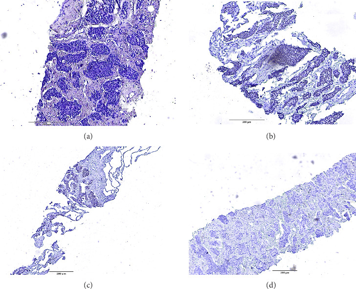
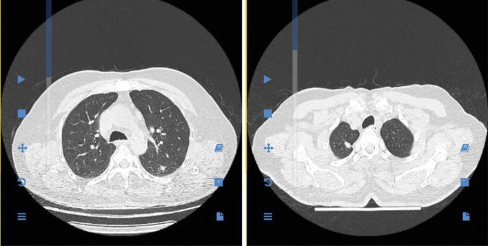
Background: Basal cell carcinoma (BCC) is the most common cutaneous malignancy, characterized by slow progression and a low propensity for metastasis. Metastatic basal cell carcinoma (mBCC) occurs in fewer than 0.1% of the cases, most frequently involving the lungs, lymph nodes, or bones. Although rare, mBCC is associated with poor prognosis and presents unique diagnostic and therapeutic challenges. Case Presentation: We report a 77-year-old male with a remote history of multiple head-and-neck BCCs, including aggressive histologic subtypes (basosquamous and micronodular), treated predominantly with Mohs surgery; the margin status varied across procedures (some tumor free and some positive). Ten years after the initial lesion, the patient developed progressive dyspnea and was found to have bilateral pulmonary nodules on chest CT. PET/CT demonstrated increased FDG uptake, and a CT-guided biopsy of the right lung nodule confirmed mBCC. There was no evidence of local recurrence at the original excision sites. p16/HPV studies were not performed on the prior cutaneous primaries. Conclusion: This case highlights the potential for delayed pulmonary metastasis in BCC, even years after apparently curative treatment. The absence of local recurrence and the bilateral lung involvement suggest hematogenous spread. Clinicians should remain vigilant for metastatic disease in patients with a history of high-risk BCC, particularly when new pulmonary symptoms arise. Imaging and immunohistochemistry are critical for diagnosis, and early detection may improve therapeutic outcomes in this rare and aggressive manifestation. In this patient, the presence of aggressive histologic subtypes and prior positive margins likely increased metastatic risk.

 求助内容:
求助内容: 应助结果提醒方式:


