Panagiotis Nikolinakos, Abhisekh Chatterjee, Efstratios Christianakis, Ioannis Alexandrou, Nikolaos Chatzikrachtis, Elisavet Kotsi, Viktor Alargkof, Ivo Donkov, Samuel Bishara, Nikolaos Zavras, Joseph M Norris
{"title":"Conservative Management of Intrascrotal Polyorchidism in a 14-Year-Old Boy: A Case Report and Review of the Current Literature.","authors":"Panagiotis Nikolinakos, Abhisekh Chatterjee, Efstratios Christianakis, Ioannis Alexandrou, Nikolaos Chatzikrachtis, Elisavet Kotsi, Viktor Alargkof, Ivo Donkov, Samuel Bishara, Nikolaos Zavras, Joseph M Norris","doi":"10.1155/criu/5258413","DOIUrl":null,"url":null,"abstract":"<p><p>Polyorchidism, or supernumerary testes (SNTs), is a rare congenital condition, management of which remains debated, particularly in paediatric cases with other concomitant features. We report a case of intrascrotal polyorchidism in a 14-year-old boy managed surgically due to parental preference and the need for histological confirmation. The patient presented with a 2-week history of painless heaviness in the scrotum. Physical examination and Doppler ultrasonography revealed a 1.8 cm mass fused to the inferior pole of the left testicle with associated Grade 1 varicocele, hydrocele and testicular appendix. Although MRI of the scrotum was initially offered, the family declined in favour of timely histological confirmation. Surgical exploration confirmed a fused supernumerary testicle and a biopsy showed normal spermatogenesis; this was consistent with Type A3 triorchidism. The patient had no complications or recurrence of symptoms at 12-month follow-up. This case highlights the use of surgical exploration in selected intrascrotal polyorchidism cases where imaging can be inconclusive or histological confirmation is required. Parental concerns and long-term reassurance may also reasonably influence management decisions.</p>","PeriodicalId":30323,"journal":{"name":"Case Reports in Urology","volume":"2025 ","pages":"5258413"},"PeriodicalIF":0.0000,"publicationDate":"2025-09-09","publicationTypes":"Journal Article","fieldsOfStudy":null,"isOpenAccess":false,"openAccessPdf":"https://www.ncbi.nlm.nih.gov/pmc/articles/PMC12440661/pdf/","citationCount":"0","resultStr":null,"platform":"Semanticscholar","paperid":null,"PeriodicalName":"Case Reports in Urology","FirstCategoryId":"1085","ListUrlMain":"https://doi.org/10.1155/criu/5258413","RegionNum":0,"RegionCategory":null,"ArticlePicture":[],"TitleCN":null,"AbstractTextCN":null,"PMCID":null,"EPubDate":"2025/1/1 0:00:00","PubModel":"eCollection","JCR":"","JCRName":"","Score":null,"Total":0}
引用次数: 0
Abstract
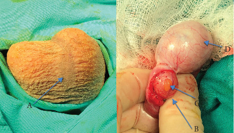
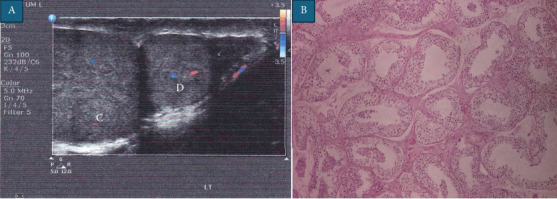
Polyorchidism, or supernumerary testes (SNTs), is a rare congenital condition, management of which remains debated, particularly in paediatric cases with other concomitant features. We report a case of intrascrotal polyorchidism in a 14-year-old boy managed surgically due to parental preference and the need for histological confirmation. The patient presented with a 2-week history of painless heaviness in the scrotum. Physical examination and Doppler ultrasonography revealed a 1.8 cm mass fused to the inferior pole of the left testicle with associated Grade 1 varicocele, hydrocele and testicular appendix. Although MRI of the scrotum was initially offered, the family declined in favour of timely histological confirmation. Surgical exploration confirmed a fused supernumerary testicle and a biopsy showed normal spermatogenesis; this was consistent with Type A3 triorchidism. The patient had no complications or recurrence of symptoms at 12-month follow-up. This case highlights the use of surgical exploration in selected intrascrotal polyorchidism cases where imaging can be inconclusive or histological confirmation is required. Parental concerns and long-term reassurance may also reasonably influence management decisions.

 求助内容:
求助内容: 应助结果提醒方式:


