Trevor Leon, Witty Kwok, Justin Lindsay, Marcelo Costa, Brittany Staarmann
{"title":"Metal artifact reduction leading to appearance of fractures in pedicle screws: illustrative case.","authors":"Trevor Leon, Witty Kwok, Justin Lindsay, Marcelo Costa, Brittany Staarmann","doi":"10.3171/CASE25358","DOIUrl":null,"url":null,"abstract":"<p><strong>Background: </strong>Metallic implants can cause imaging artifacts on CT and MRI. Although metal artifact reduction (MAR) techniques have enhanced imaging clarity, they can also lead to distortions that may resemble complications such as hardware fractures. The false appearance of fractured hardware, or \"pseudofractures,\" can distort imaging interpretation and result in unnecessary revision surgery.</p><p><strong>Observations: </strong>A 65-year-old male sustained a T12-L1 fracture extending through the disc space with ligamentous interruption after a bicycle accident. The patient underwent T11-L2 posterior fusion with pedicle screws and was discharged in a thoracolumbar sacral orthosis brace after an uncomplicated postoperative course. Concerns about brace compliance prompted imaging, which demonstrated screw fractures and led to discussion of revision procedure. Repeat imaging revealed that these were artifacts, not hardware failure. No revision was needed and the patient continued to recover.</p><p><strong>Lessons: </strong>This case emphasizes the importance of understanding imaging techniques that may impact interpretation and implications for surgical decision-making. Imaging artifacts should be considered when hardware fractures exist at multiple levels without suggestive history of new symptoms. Obtaining plain radiographs or images without MAR may assist in diagnostic uncertainty. Discussing MAR use with the radiology department at imaging acquisition may improve both imaging interpretations and resource utilization. https://thejns.org/doi/10.3171/CASE25358.</p>","PeriodicalId":94098,"journal":{"name":"Journal of neurosurgery. Case lessons","volume":"10 11","pages":""},"PeriodicalIF":0.0000,"publicationDate":"2025-09-15","publicationTypes":"Journal Article","fieldsOfStudy":null,"isOpenAccess":false,"openAccessPdf":"https://www.ncbi.nlm.nih.gov/pmc/articles/PMC12435379/pdf/","citationCount":"0","resultStr":null,"platform":"Semanticscholar","paperid":null,"PeriodicalName":"Journal of neurosurgery. Case lessons","FirstCategoryId":"1085","ListUrlMain":"https://doi.org/10.3171/CASE25358","RegionNum":0,"RegionCategory":null,"ArticlePicture":[],"TitleCN":null,"AbstractTextCN":null,"PMCID":null,"EPubDate":"","PubModel":"","JCR":"","JCRName":"","Score":null,"Total":0}
引用次数: 0
Abstract
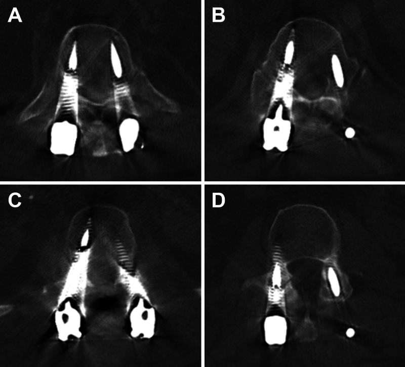
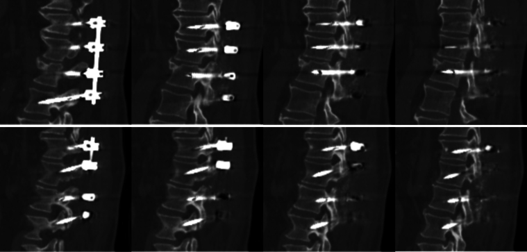
Background: Metallic implants can cause imaging artifacts on CT and MRI. Although metal artifact reduction (MAR) techniques have enhanced imaging clarity, they can also lead to distortions that may resemble complications such as hardware fractures. The false appearance of fractured hardware, or "pseudofractures," can distort imaging interpretation and result in unnecessary revision surgery.
Observations: A 65-year-old male sustained a T12-L1 fracture extending through the disc space with ligamentous interruption after a bicycle accident. The patient underwent T11-L2 posterior fusion with pedicle screws and was discharged in a thoracolumbar sacral orthosis brace after an uncomplicated postoperative course. Concerns about brace compliance prompted imaging, which demonstrated screw fractures and led to discussion of revision procedure. Repeat imaging revealed that these were artifacts, not hardware failure. No revision was needed and the patient continued to recover.
Lessons: This case emphasizes the importance of understanding imaging techniques that may impact interpretation and implications for surgical decision-making. Imaging artifacts should be considered when hardware fractures exist at multiple levels without suggestive history of new symptoms. Obtaining plain radiographs or images without MAR may assist in diagnostic uncertainty. Discussing MAR use with the radiology department at imaging acquisition may improve both imaging interpretations and resource utilization. https://thejns.org/doi/10.3171/CASE25358.


 求助内容:
求助内容: 应助结果提醒方式:


