Hughes W Benjamin, Lauren E Corliss, Taylor N Murray, Gregory Chamberlin, Deveney Franklin, Mark A Attiah
{"title":"Extraskeletal para-articular osteochondroma adjacent to the cervical spine: illustrative case.","authors":"Hughes W Benjamin, Lauren E Corliss, Taylor N Murray, Gregory Chamberlin, Deveney Franklin, Mark A Attiah","doi":"10.3171/CASE25468","DOIUrl":null,"url":null,"abstract":"<p><strong>Background: </strong>Extraskeletal osteochondromas are well-circumscribed osteocartilaginous lesions arising from soft tissues without bone continuity. Extraskeletal osteochondromas may present in a para-articular location, although few reported cases have occurred near the spine. Clinical diagnosis remains challenging as these tumors can be difficult to distinguish from other ossified soft tissue lesions. Treatment includes management by observation or resection. Here the authors present a rare case of an extraskeletal osteochondroma near the cervical spine.</p><p><strong>Observations: </strong>A 56-year-old male presented with a palpable left paraspinal suboccipital mass that had slowly progressed over 5 years. The patient was asymptomatic and the neurological examination was nonfocal. MRI revealed an approximately 6-cm well-circumscribed heterogeneous mass in the left suboccipital area that lacked direct contact with the spine or calvarium. A hypointense capsule on T2-weighted MRI and multiple hypointense septations on T1- and T2-weighted MRI were identified. The slow rate of tumor growth suggested a benign tumor. CT imaging of the chest, abdomen, and pelvis was obtained to rule out malignancy. Surgical intervention was offered and accepted by the patient. At 2 months postoperatively, imaging demonstrated no recurrence, and the patient was asymptomatic and had improved range of motion.</p><p><strong>Lessons: </strong>Extraskeletal osteochondroma should be considered when diagnosing osteocartilaginous paraspinal masses. https://thejns.org/doi/10.3171/CASE25468.</p>","PeriodicalId":94098,"journal":{"name":"Journal of neurosurgery. Case lessons","volume":"10 11","pages":""},"PeriodicalIF":0.0000,"publicationDate":"2025-09-15","publicationTypes":"Journal Article","fieldsOfStudy":null,"isOpenAccess":false,"openAccessPdf":"https://www.ncbi.nlm.nih.gov/pmc/articles/PMC12435374/pdf/","citationCount":"0","resultStr":null,"platform":"Semanticscholar","paperid":null,"PeriodicalName":"Journal of neurosurgery. Case lessons","FirstCategoryId":"1085","ListUrlMain":"https://doi.org/10.3171/CASE25468","RegionNum":0,"RegionCategory":null,"ArticlePicture":[],"TitleCN":null,"AbstractTextCN":null,"PMCID":null,"EPubDate":"","PubModel":"","JCR":"","JCRName":"","Score":null,"Total":0}
引用次数: 0
Abstract
Background: Extraskeletal osteochondromas are well-circumscribed osteocartilaginous lesions arising from soft tissues without bone continuity. Extraskeletal osteochondromas may present in a para-articular location, although few reported cases have occurred near the spine. Clinical diagnosis remains challenging as these tumors can be difficult to distinguish from other ossified soft tissue lesions. Treatment includes management by observation or resection. Here the authors present a rare case of an extraskeletal osteochondroma near the cervical spine.
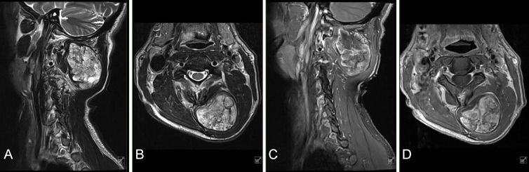
Observations: A 56-year-old male presented with a palpable left paraspinal suboccipital mass that had slowly progressed over 5 years. The patient was asymptomatic and the neurological examination was nonfocal. MRI revealed an approximately 6-cm well-circumscribed heterogeneous mass in the left suboccipital area that lacked direct contact with the spine or calvarium. A hypointense capsule on T2-weighted MRI and multiple hypointense septations on T1- and T2-weighted MRI were identified. The slow rate of tumor growth suggested a benign tumor. CT imaging of the chest, abdomen, and pelvis was obtained to rule out malignancy. Surgical intervention was offered and accepted by the patient. At 2 months postoperatively, imaging demonstrated no recurrence, and the patient was asymptomatic and had improved range of motion.
Lessons: Extraskeletal osteochondroma should be considered when diagnosing osteocartilaginous paraspinal masses. https://thejns.org/doi/10.3171/CASE25468.

 求助内容:
求助内容: 应助结果提醒方式:


