Sarah Campos, Firooz Salami, Qiuyue Chen, Cornelia Putz, Stefanos Tsitlakidis, Sebastian I Wolf
{"title":"The Heidelberg Functional Foot Model-Application to Cavovarus and Equinovarus Feet.","authors":"Sarah Campos, Firooz Salami, Qiuyue Chen, Cornelia Putz, Stefanos Tsitlakidis, Sebastian I Wolf","doi":"10.1002/jfa2.70085","DOIUrl":null,"url":null,"abstract":"<p><p>Multisegment foot models have become increasingly important in biomechanical research and clinical gait analysis but often face limitations in defining joint positions. Often, they rely on simplified methods, such as using the midpoint between two markers to represent a joint, which lacks functional verification. In contrast, phenomenological angles, such as the medial arch angle, bypass joint center calculations, and offer sensitive, radiologically aligned indicators of foot mechanics. The Heidelberg functional foot model (HFFM) integrates functionally verified joint positions in combination with clinically relevant phenomenological measures, thereby enhancing clinical interpretability in gait analysis. The marker placement of the HFFM is based on the Heidelberg foot measurement method (HFMM). A four-segment model (shank, hindfoot, forefoot, and hallux) is defined. Anatomical coordinate systems are established via regression formulas derived from functional joint parameter determination. Kinematic angles are compared with radiological measures. Additionally, six clinically relevant angles of the HFMM are integrated into the HFFM. The method is applied to cavovarus (CV, 19 feet), equinovarus (EV, 31 feet), and typically developed feet (TD, 88 feet). EV feet show more pronounced hindfoot varus and forefoot adduction than CV and TD feet. Within the parameters adopted from the HFMM, EV feet exhibit increased subtalar inversion and a stronger medial arch than CV. Significant correlations are identified between hindfoot/shank flexion, forefoot/hindfoot flexion and medial arch, and radiological angles. The HFFM is sensitive for analyzing equinvarus and cavovarus deformities without applying static offsets due to the functional approach. It enables calculating kinetics to better understand the biomechanics of foot deformities.</p>","PeriodicalId":49164,"journal":{"name":"Journal of Foot and Ankle Research","volume":"18 3","pages":"e70085"},"PeriodicalIF":2.2000,"publicationDate":"2025-09-01","publicationTypes":"Journal Article","fieldsOfStudy":null,"isOpenAccess":false,"openAccessPdf":"https://www.ncbi.nlm.nih.gov/pmc/articles/PMC12436176/pdf/","citationCount":"0","resultStr":null,"platform":"Semanticscholar","paperid":null,"PeriodicalName":"Journal of Foot and Ankle Research","FirstCategoryId":"3","ListUrlMain":"https://doi.org/10.1002/jfa2.70085","RegionNum":3,"RegionCategory":"医学","ArticlePicture":[],"TitleCN":null,"AbstractTextCN":null,"PMCID":null,"EPubDate":"","PubModel":"","JCR":"Q1","JCRName":"ORTHOPEDICS","Score":null,"Total":0}
引用次数: 0
Abstract
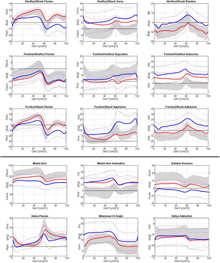
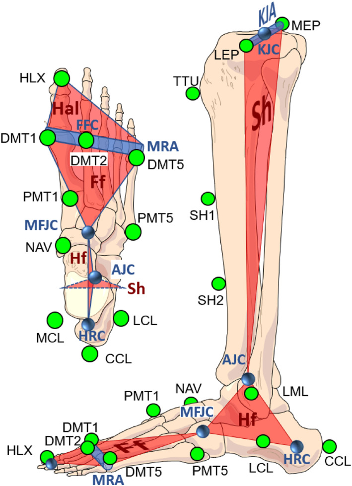
Multisegment foot models have become increasingly important in biomechanical research and clinical gait analysis but often face limitations in defining joint positions. Often, they rely on simplified methods, such as using the midpoint between two markers to represent a joint, which lacks functional verification. In contrast, phenomenological angles, such as the medial arch angle, bypass joint center calculations, and offer sensitive, radiologically aligned indicators of foot mechanics. The Heidelberg functional foot model (HFFM) integrates functionally verified joint positions in combination with clinically relevant phenomenological measures, thereby enhancing clinical interpretability in gait analysis. The marker placement of the HFFM is based on the Heidelberg foot measurement method (HFMM). A four-segment model (shank, hindfoot, forefoot, and hallux) is defined. Anatomical coordinate systems are established via regression formulas derived from functional joint parameter determination. Kinematic angles are compared with radiological measures. Additionally, six clinically relevant angles of the HFMM are integrated into the HFFM. The method is applied to cavovarus (CV, 19 feet), equinovarus (EV, 31 feet), and typically developed feet (TD, 88 feet). EV feet show more pronounced hindfoot varus and forefoot adduction than CV and TD feet. Within the parameters adopted from the HFMM, EV feet exhibit increased subtalar inversion and a stronger medial arch than CV. Significant correlations are identified between hindfoot/shank flexion, forefoot/hindfoot flexion and medial arch, and radiological angles. The HFFM is sensitive for analyzing equinvarus and cavovarus deformities without applying static offsets due to the functional approach. It enables calculating kinetics to better understand the biomechanics of foot deformities.
期刊介绍:
Journal of Foot and Ankle Research, the official journal of the Australian Podiatry Association and The College of Podiatry (UK), is an open access journal that encompasses all aspects of policy, organisation, delivery and clinical practice related to the assessment, diagnosis, prevention and management of foot and ankle disorders.
Journal of Foot and Ankle Research covers a wide range of clinical subject areas, including diabetology, paediatrics, sports medicine, gerontology and geriatrics, foot surgery, physical therapy, dermatology, wound management, radiology, biomechanics and bioengineering, orthotics and prosthetics, as well the broad areas of epidemiology, policy, organisation and delivery of services related to foot and ankle care.
The journal encourages submissions from all health professionals who manage lower limb conditions, including podiatrists, nurses, physical therapists and physiotherapists, orthopaedists, manual therapists, medical specialists and general medical practitioners, as well as health service researchers concerned with foot and ankle care.
The Australian Podiatry Association and the College of Podiatry (UK) have reserve funds to cover the article-processing charge for manuscripts submitted by its members. Society members can email the appropriate contact at Australian Podiatry Association or The College of Podiatry to obtain the corresponding code to enter on submission.
 求助内容:
求助内容: 应助结果提醒方式:


