Cortical changes in women with systemic lupus erythematosus with mild cognitive impairment: a voxel-based morphometry and surface-based morphometry study.
{"title":"Cortical changes in women with systemic lupus erythematosus with mild cognitive impairment: a voxel-based morphometry and surface-based morphometry study.","authors":"Minghuang Mo, Yifan Yang, Shuang Liu, Ru Bai, Shu Li, Ruotong Zhao, Xinyu Xu, Yuqi Cheng, Jian Xu","doi":"10.1136/lupus-2025-001523","DOIUrl":null,"url":null,"abstract":"<p><strong>Background: </strong>The purpose of this study was to reveal the morphological changes of grey matter (GM) in women systemic lupus erythematosus (wSLE) patients with mild cognitive impairment (MCI) with normal conventional MRI.</p><p><strong>Methods: </strong>The differences in brain morphological indicators among wSLE with MCI, wSLE without MCI and women healthy control (wHC) group were calculated and compared by voxel-based morphometry and surface-based morphometry. The GM volume (GMV), cortical thickness (CT), indicators of cortical complexity, including fractal dimension (FD), gyration index (GI), sulcus depth, the relationship between brain morphological indicators and clinical features, were analysed.</p><p><strong>Results: </strong>In comparison to wSLE patients without MCI (n=36), wSLE with MCI (n=26) demonstrated a significant decrease in FD of the left lateral orbitofrontal gyrus. When compared with the wHC group (n=36), both wSLE patients with MCI and wSLE without MCI group exhibited a reduction in GMV in the medial of right superior frontal gyrus, a thinning of CT in the left paracentral and postcentral gyrus as well as in the right pars triangularis gyrus and superior frontal gyrus. Within the wSLE group, Mini-Mental State Examination scores were positively correlated with GMV in the middle of right superior frontal gyrus and with the FD of the left lateral orbitofrontal gyrus.</p><p><strong>Conclusion: </strong>WSLE patients with MCI have brain morphological changes such as reduced GMV, thinning CT, reduced FD and increased GI. Cortical morphological changes may be involved in the pathological process of MCI in wSLE patients.</p>","PeriodicalId":18126,"journal":{"name":"Lupus Science & Medicine","volume":"12 2","pages":""},"PeriodicalIF":3.5000,"publicationDate":"2025-09-14","publicationTypes":"Journal Article","fieldsOfStudy":null,"isOpenAccess":false,"openAccessPdf":"https://www.ncbi.nlm.nih.gov/pmc/articles/PMC12434781/pdf/","citationCount":"0","resultStr":null,"platform":"Semanticscholar","paperid":null,"PeriodicalName":"Lupus Science & Medicine","FirstCategoryId":"3","ListUrlMain":"https://doi.org/10.1136/lupus-2025-001523","RegionNum":2,"RegionCategory":"医学","ArticlePicture":[],"TitleCN":null,"AbstractTextCN":null,"PMCID":null,"EPubDate":"","PubModel":"","JCR":"Q1","JCRName":"RHEUMATOLOGY","Score":null,"Total":0}
引用次数: 0
Abstract
Background: The purpose of this study was to reveal the morphological changes of grey matter (GM) in women systemic lupus erythematosus (wSLE) patients with mild cognitive impairment (MCI) with normal conventional MRI.
Methods: The differences in brain morphological indicators among wSLE with MCI, wSLE without MCI and women healthy control (wHC) group were calculated and compared by voxel-based morphometry and surface-based morphometry. The GM volume (GMV), cortical thickness (CT), indicators of cortical complexity, including fractal dimension (FD), gyration index (GI), sulcus depth, the relationship between brain morphological indicators and clinical features, were analysed.
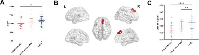
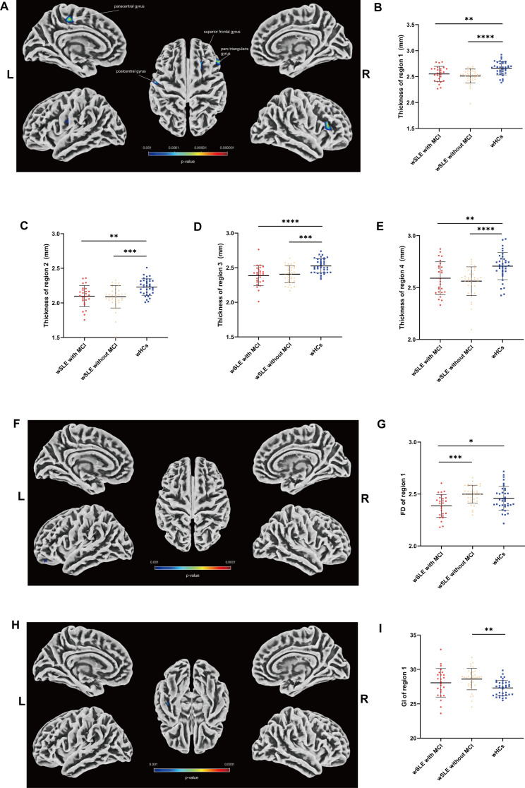
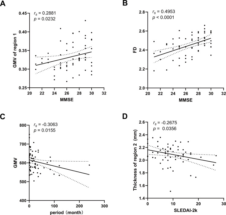
Results: In comparison to wSLE patients without MCI (n=36), wSLE with MCI (n=26) demonstrated a significant decrease in FD of the left lateral orbitofrontal gyrus. When compared with the wHC group (n=36), both wSLE patients with MCI and wSLE without MCI group exhibited a reduction in GMV in the medial of right superior frontal gyrus, a thinning of CT in the left paracentral and postcentral gyrus as well as in the right pars triangularis gyrus and superior frontal gyrus. Within the wSLE group, Mini-Mental State Examination scores were positively correlated with GMV in the middle of right superior frontal gyrus and with the FD of the left lateral orbitofrontal gyrus.
Conclusion: WSLE patients with MCI have brain morphological changes such as reduced GMV, thinning CT, reduced FD and increased GI. Cortical morphological changes may be involved in the pathological process of MCI in wSLE patients.
期刊介绍:
Lupus Science & Medicine is a global, peer reviewed, open access online journal that provides a central point for publication of basic, clinical, translational, and epidemiological studies of all aspects of lupus and related diseases. It is the first lupus-specific open access journal in the world and was developed in response to the need for a barrier-free forum for publication of groundbreaking studies in lupus. The journal publishes research on lupus from fields including, but not limited to: rheumatology, dermatology, nephrology, immunology, pediatrics, cardiology, hepatology, pulmonology, obstetrics and gynecology, and psychiatry.
 求助内容:
求助内容: 应助结果提醒方式:


