Emmanuelle D Aiyegbusi, James P Reynolds, Ross O'Carroll, Ruth Colbert, Christopher Carew, Dearbhaile Dooley
{"title":"Forced polarisation of microglia by IL-13 is modified by inflammatory and microenvironmental context.","authors":"Emmanuelle D Aiyegbusi, James P Reynolds, Ross O'Carroll, Ruth Colbert, Christopher Carew, Dearbhaile Dooley","doi":"10.1007/s00011-025-02089-2","DOIUrl":null,"url":null,"abstract":"<p><strong>Background: </strong>Traumatic spinal cord injury (SCI) is a severe clinical challenge, often leading to long-term sensory, motor, and autonomic dysfunction. The SCI cascade involves a primary physical damage phase, followed by a secondary phase of inflammatory signalling driven by microglia and other infiltrating immune cells. Immunomodulatory therapies may help promote healing and restrict secondary damage. We have previously demonstrated that interleukin (IL)-13 delivery improves functional and histopathological recovery after SCI in murine models, primarily by polarising macrophages towards an alternatively activated pro-reparative M2-like phenotype and reducing axonal contacts. Although microglia respond robustly to IL-13 in vitro, polarisation of microglia in vivo is more difficult. To better understand what conditions may restrict microglial responses to IL-13 in vivo, we sought to examine the effect of cellular context or microenvironment on IL-13 efficacy in forcing microglia polarisation in vitro.</p><p><strong>Methods: </strong>BV2 and murine induced pluripotent stem cell (miPSC)-derived microglia were treated with IL-13 alone or in combination with lipopolysaccharide (LPS), acidic media, extracellular matrix components, high glutamate or high potassium concentrations. Following this phenotypic changes including morphology, gene/protein expression (TNFα, IL-1β, iNOS, Arg-1, CD206, F4-80) and cytokine release (TNFα) were measured using high-content screening, RT-qPCR, immunohistochemistry, and ELISA.</p><p><strong>Results: </strong>IL-13 leads to increased expression of the anti-inflammatory marker Arg-1 while lowering expression and secretion of the pro-inflammatory markers IL-1β, iNOS, and TNFα, and expression of the microglia activation marker F4-80, signifying effective polarisation of microglia. Concomitant administration of LPS with IL-13 reduces IL-13 polarisation efficacy in microglia. Forced polarisation of microglia is also compromised by high glutamate tone, acidosis, hyperkalemia, and extracellular fibronectin, suggesting microenvironmental contexts seen in neurotrauma directly act on microglia to limit polarisation potential.</p><p><strong>Conclusions: </strong>Our study demonstrates that the post-SCI environment dampens IL-13 efficacy on microglia. Taken together these data caution against simple immunomodulatory strategies and suggest that effective polarisation of microglia in vivo will require multimodal approaches.</p>","PeriodicalId":13550,"journal":{"name":"Inflammation Research","volume":"74 1","pages":"130"},"PeriodicalIF":5.4000,"publicationDate":"2025-09-16","publicationTypes":"Journal Article","fieldsOfStudy":null,"isOpenAccess":false,"openAccessPdf":"https://www.ncbi.nlm.nih.gov/pmc/articles/PMC12441057/pdf/","citationCount":"0","resultStr":null,"platform":"Semanticscholar","paperid":null,"PeriodicalName":"Inflammation Research","FirstCategoryId":"3","ListUrlMain":"https://doi.org/10.1007/s00011-025-02089-2","RegionNum":3,"RegionCategory":"医学","ArticlePicture":[],"TitleCN":null,"AbstractTextCN":null,"PMCID":null,"EPubDate":"","PubModel":"","JCR":"Q2","JCRName":"CELL BIOLOGY","Score":null,"Total":0}
引用次数: 0
Abstract
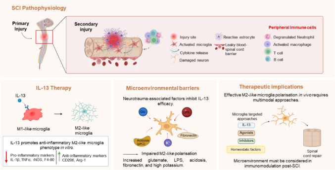
Background: Traumatic spinal cord injury (SCI) is a severe clinical challenge, often leading to long-term sensory, motor, and autonomic dysfunction. The SCI cascade involves a primary physical damage phase, followed by a secondary phase of inflammatory signalling driven by microglia and other infiltrating immune cells. Immunomodulatory therapies may help promote healing and restrict secondary damage. We have previously demonstrated that interleukin (IL)-13 delivery improves functional and histopathological recovery after SCI in murine models, primarily by polarising macrophages towards an alternatively activated pro-reparative M2-like phenotype and reducing axonal contacts. Although microglia respond robustly to IL-13 in vitro, polarisation of microglia in vivo is more difficult. To better understand what conditions may restrict microglial responses to IL-13 in vivo, we sought to examine the effect of cellular context or microenvironment on IL-13 efficacy in forcing microglia polarisation in vitro.
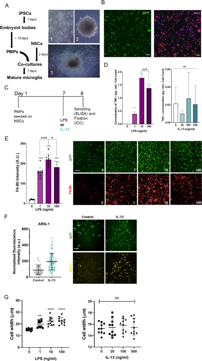
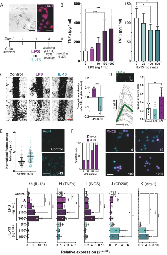
Methods: BV2 and murine induced pluripotent stem cell (miPSC)-derived microglia were treated with IL-13 alone or in combination with lipopolysaccharide (LPS), acidic media, extracellular matrix components, high glutamate or high potassium concentrations. Following this phenotypic changes including morphology, gene/protein expression (TNFα, IL-1β, iNOS, Arg-1, CD206, F4-80) and cytokine release (TNFα) were measured using high-content screening, RT-qPCR, immunohistochemistry, and ELISA.
Results: IL-13 leads to increased expression of the anti-inflammatory marker Arg-1 while lowering expression and secretion of the pro-inflammatory markers IL-1β, iNOS, and TNFα, and expression of the microglia activation marker F4-80, signifying effective polarisation of microglia. Concomitant administration of LPS with IL-13 reduces IL-13 polarisation efficacy in microglia. Forced polarisation of microglia is also compromised by high glutamate tone, acidosis, hyperkalemia, and extracellular fibronectin, suggesting microenvironmental contexts seen in neurotrauma directly act on microglia to limit polarisation potential.
Conclusions: Our study demonstrates that the post-SCI environment dampens IL-13 efficacy on microglia. Taken together these data caution against simple immunomodulatory strategies and suggest that effective polarisation of microglia in vivo will require multimodal approaches.
期刊介绍:
Inflammation Research (IR) publishes peer-reviewed papers on all aspects of inflammation and related fields including histopathology, immunological mechanisms, gene expression, mediators, experimental models, clinical investigations and the effect of drugs. Related fields are broadly defined and include for instance, allergy and asthma, shock, pain, joint damage, skin disease as well as clinical trials of relevant drugs.
 求助内容:
求助内容: 应助结果提醒方式:


