Yang Shan Edmond Lim, Shuyi Guo, Zhuyi Rebekah Lee, Emma Choon Hwee Lee, Timothy Shao Ern Tan
{"title":"A visual compendium of teratomas and their diverse anatomical locations.","authors":"Yang Shan Edmond Lim, Shuyi Guo, Zhuyi Rebekah Lee, Emma Choon Hwee Lee, Timothy Shao Ern Tan","doi":"10.1177/20584601251377911","DOIUrl":null,"url":null,"abstract":"<p><p>Teratomas are a common type of germ cell tumours which may be benign or malignant. Benign mature teratomas are the most frequent subtype and typically show intralesional fat and calcifications within a cystic mass. Immature/malignant teratomas are usually larger with irregular solid components, coarse calcifications, small amounts of fat, and with or without necrosis or haemorrhage. Teratomas can manifest in various anatomical locations, particularly in the sacrococcygeal, gonadal, mediastinal, retroperitoneal, and intracranial regions. This article explores the imaging characteristics and diverse locations of teratomas as well as discusses about possible differential diagnoses to facilitate early detection and ensure prompt treatment.</p>","PeriodicalId":72063,"journal":{"name":"Acta radiologica open","volume":"14 9","pages":"20584601251377911"},"PeriodicalIF":1.0000,"publicationDate":"2025-09-10","publicationTypes":"Journal Article","fieldsOfStudy":null,"isOpenAccess":false,"openAccessPdf":"https://www.ncbi.nlm.nih.gov/pmc/articles/PMC12423518/pdf/","citationCount":"0","resultStr":null,"platform":"Semanticscholar","paperid":null,"PeriodicalName":"Acta radiologica open","FirstCategoryId":"1085","ListUrlMain":"https://doi.org/10.1177/20584601251377911","RegionNum":0,"RegionCategory":null,"ArticlePicture":[],"TitleCN":null,"AbstractTextCN":null,"PMCID":null,"EPubDate":"2025/9/1 0:00:00","PubModel":"eCollection","JCR":"Q4","JCRName":"RADIOLOGY, NUCLEAR MEDICINE & MEDICAL IMAGING","Score":null,"Total":0}
引用次数: 0
Abstract
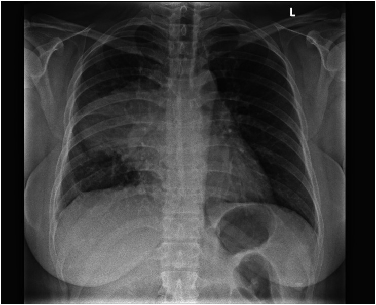
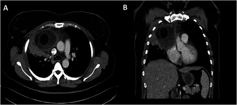
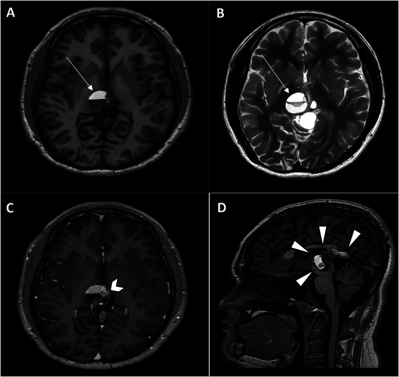
Teratomas are a common type of germ cell tumours which may be benign or malignant. Benign mature teratomas are the most frequent subtype and typically show intralesional fat and calcifications within a cystic mass. Immature/malignant teratomas are usually larger with irregular solid components, coarse calcifications, small amounts of fat, and with or without necrosis or haemorrhage. Teratomas can manifest in various anatomical locations, particularly in the sacrococcygeal, gonadal, mediastinal, retroperitoneal, and intracranial regions. This article explores the imaging characteristics and diverse locations of teratomas as well as discusses about possible differential diagnoses to facilitate early detection and ensure prompt treatment.

 求助内容:
求助内容: 应助结果提醒方式:


