{"title":"Analysis of the clinical value of gemstone spectral computed tomography imaging in the preoperative assessment of colorectal cancer.","authors":"Wei Liu, De-Min Kong, Jian-Kun An, Li-Tao Song","doi":"10.4240/wjgs.v17.i8.105391","DOIUrl":null,"url":null,"abstract":"<p><strong>Background: </strong>The diagnostic accuracy for detecting metastatic lymph nodes in colorectal cancer (CRC) remains suboptimal. To address this limitation, our study investigates the potential of gemstone spectral computed tomography imaging (GSI) to improve diagnostic accuracy in lymph node metastasis (LNM) assessment.</p><p><strong>Aim: </strong>To extensively investigate the clinical utility of GSI in the preoperative assessment of CRC.</p><p><strong>Methods: </strong>The subject population included 200 patients with CRC who were admitted to Zibo Central Hospital from January 2022 to December 2023. All patients underwent dual-phase contrast-enhanced scans in the arterial and venous phases using GSI before surgical intervention. During the research, meticulous quantification was conducted regarding the number of patients with CRC with LNM as well as the exact count of metastatic lymph nodes. Moreover, for both metastatic and non-metastatic lymph nodes, the short diameter at the maximum cross-sectional area (covering the axial, sagittal, and coronal planes), morphological features (including manifestations such as margin blurring, aggregation, and enhancement), and spectral parameters in the arterial and venous phases [specifically iodine concentration (IC), normalized IC (NIC), and the slope of the spectral curve (λ<sub>HU</sub>)] were measured and recorded, and a comparative analysis was conducted. The diagnostic efficacy of each index with differences was systematically assessed using the receiver operating characteristic (ROC) curve. Concurrently, receiver operating characteristic curves were constructed for LNM screening based on the short diameter at the maximum cross-sectional area of lymph nodes and each spectral parameter in the arterial and venous phases.</p><p><strong>Results: </strong>The area under the curve of GSI for diagnosing LNM in patients with CRC can reach 0.897, with sensitivity, specificity, and accuracy of 92.59%, 85.87%, and 89.50%, respectively. A total of 265 lymph nodes were analyzed from the 200 participants with CRC, with metastatic lymph nodes accounting for 56.60%. Compared with non-metastatic lymph nodes, the short diameters of metastatic lymph nodes in the axial, sagittal, and coronal planes were significantly increased, whereas the IC values in the arterial and venous phases, the NIC value in the arterial phase, and the λ<sub>HU</sub> values in the arterial and venous phases were significantly decreased. The short axial, sagittal, and coronal diameters, arterial-phase IC, venous-phase IC, arterial-phase NIC, arterial-phase λ<sub>HU</sub>, and venous-phase λ<sub>HU</sub> for diagnosing metastatic lymph nodes demonstrated area under the curve values of 0.631, 0.681, 0.659, 0.862, 0.808, 0.831, 0.801, and 0.706, respectively.</p><p><strong>Conclusion: </strong>GSI exhibits substantial clinical significance in the preoperative assessment of CRC. Among the parameters assessed, the arterial-phase IC demonstrates the most outstanding diagnostic performance, effectively improving the diagnostic efficacy for preoperative LNM in CRC.</p>","PeriodicalId":23759,"journal":{"name":"World Journal of Gastrointestinal Surgery","volume":"17 8","pages":"105391"},"PeriodicalIF":1.7000,"publicationDate":"2025-08-27","publicationTypes":"Journal Article","fieldsOfStudy":null,"isOpenAccess":false,"openAccessPdf":"https://www.ncbi.nlm.nih.gov/pmc/articles/PMC12432544/pdf/","citationCount":"0","resultStr":null,"platform":"Semanticscholar","paperid":null,"PeriodicalName":"World Journal of Gastrointestinal Surgery","FirstCategoryId":"3","ListUrlMain":"https://doi.org/10.4240/wjgs.v17.i8.105391","RegionNum":4,"RegionCategory":"医学","ArticlePicture":[],"TitleCN":null,"AbstractTextCN":null,"PMCID":null,"EPubDate":"","PubModel":"","JCR":"Q3","JCRName":"GASTROENTEROLOGY & HEPATOLOGY","Score":null,"Total":0}
引用次数: 0
Abstract
Background: The diagnostic accuracy for detecting metastatic lymph nodes in colorectal cancer (CRC) remains suboptimal. To address this limitation, our study investigates the potential of gemstone spectral computed tomography imaging (GSI) to improve diagnostic accuracy in lymph node metastasis (LNM) assessment.
Aim: To extensively investigate the clinical utility of GSI in the preoperative assessment of CRC.
Methods: The subject population included 200 patients with CRC who were admitted to Zibo Central Hospital from January 2022 to December 2023. All patients underwent dual-phase contrast-enhanced scans in the arterial and venous phases using GSI before surgical intervention. During the research, meticulous quantification was conducted regarding the number of patients with CRC with LNM as well as the exact count of metastatic lymph nodes. Moreover, for both metastatic and non-metastatic lymph nodes, the short diameter at the maximum cross-sectional area (covering the axial, sagittal, and coronal planes), morphological features (including manifestations such as margin blurring, aggregation, and enhancement), and spectral parameters in the arterial and venous phases [specifically iodine concentration (IC), normalized IC (NIC), and the slope of the spectral curve (λHU)] were measured and recorded, and a comparative analysis was conducted. The diagnostic efficacy of each index with differences was systematically assessed using the receiver operating characteristic (ROC) curve. Concurrently, receiver operating characteristic curves were constructed for LNM screening based on the short diameter at the maximum cross-sectional area of lymph nodes and each spectral parameter in the arterial and venous phases.
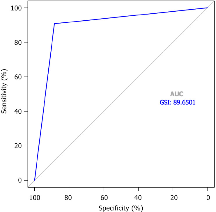
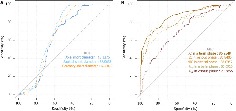
Results: The area under the curve of GSI for diagnosing LNM in patients with CRC can reach 0.897, with sensitivity, specificity, and accuracy of 92.59%, 85.87%, and 89.50%, respectively. A total of 265 lymph nodes were analyzed from the 200 participants with CRC, with metastatic lymph nodes accounting for 56.60%. Compared with non-metastatic lymph nodes, the short diameters of metastatic lymph nodes in the axial, sagittal, and coronal planes were significantly increased, whereas the IC values in the arterial and venous phases, the NIC value in the arterial phase, and the λHU values in the arterial and venous phases were significantly decreased. The short axial, sagittal, and coronal diameters, arterial-phase IC, venous-phase IC, arterial-phase NIC, arterial-phase λHU, and venous-phase λHU for diagnosing metastatic lymph nodes demonstrated area under the curve values of 0.631, 0.681, 0.659, 0.862, 0.808, 0.831, 0.801, and 0.706, respectively.
Conclusion: GSI exhibits substantial clinical significance in the preoperative assessment of CRC. Among the parameters assessed, the arterial-phase IC demonstrates the most outstanding diagnostic performance, effectively improving the diagnostic efficacy for preoperative LNM in CRC.

 求助内容:
求助内容: 应助结果提醒方式:


