{"title":"Effects of low-density lipoprotein cholesterol on lymph node metastasis after radical esophagectomy.","authors":"Xin-Jian Xu, Shi-Wei Liu, Jia-Qi Li, Ming He, Hui Wang, Qing-Ju Meng","doi":"10.4240/wjgs.v17.i8.106898","DOIUrl":null,"url":null,"abstract":"<p><strong>Background: </strong>Esophageal cancer (EC) is one of the most common malignancies worldwide, and lymph node (LN) metastasis remains one of the leading causes of EC recurrence. Metabolic disorders critically affect cancer progression, and lipid levels are closely associated with the occurrence of EC and several other tumor types. This study analyzed pretreatment lipid levels to determine their association with LN metastasis.</p><p><strong>Aim: </strong>To dissect the possible mechanisms underlying LN metastasis and clarify the prognostic role of lipid profiles in EC.</p><p><strong>Methods: </strong>Serum lipid levels and clinicopathological information were retrospectively collected from 294 patients, and risk factors for LN metastasis were confirmed using a logistic regression model. Latent factors were explored using information from publicly accessible databases and immunofluorescence and immunohistochemical staining techniques.</p><p><strong>Results: </strong>High serum levels of low-density lipoprotein (LDL) cholesterol promote LN metastasis in EC, while high-density lipoprotein cholesterol has the opposite role. Information of a public database revealed that LDL receptors LRP5 and LRP6 are highly expressed in ECs, and LRP6 overexpression positively correlated with the infiltration of B lymphocytes and a poor prognosis. Immunofluorescence and immunohistochemical staining revealed that the expression of LRP6 and infiltrated B lymphocytes in patients with ≥ 1 regional LN metastasis, containing N1-3 (N+ group) were significantly higher than those in the N0 group. LRP6 was also highly expressed in the B lymphocytes of the N+ group. There was no difference in CXCL13 expression between the N+ and N0 groups. However, CXCR5 expression was significantly higher in the N0 group than in the N+ group.</p><p><strong>Conclusion: </strong>High serum LDL levels can promote LN metastasis in EC, and the mechanisms may be related to LRP6 expression and the infiltration of B lymphocytes.</p>","PeriodicalId":23759,"journal":{"name":"World Journal of Gastrointestinal Surgery","volume":"17 8","pages":"106898"},"PeriodicalIF":1.7000,"publicationDate":"2025-08-27","publicationTypes":"Journal Article","fieldsOfStudy":null,"isOpenAccess":false,"openAccessPdf":"https://www.ncbi.nlm.nih.gov/pmc/articles/PMC12427011/pdf/","citationCount":"0","resultStr":null,"platform":"Semanticscholar","paperid":null,"PeriodicalName":"World Journal of Gastrointestinal Surgery","FirstCategoryId":"3","ListUrlMain":"https://doi.org/10.4240/wjgs.v17.i8.106898","RegionNum":4,"RegionCategory":"医学","ArticlePicture":[],"TitleCN":null,"AbstractTextCN":null,"PMCID":null,"EPubDate":"","PubModel":"","JCR":"Q3","JCRName":"GASTROENTEROLOGY & HEPATOLOGY","Score":null,"Total":0}
引用次数: 0
Abstract
Background: Esophageal cancer (EC) is one of the most common malignancies worldwide, and lymph node (LN) metastasis remains one of the leading causes of EC recurrence. Metabolic disorders critically affect cancer progression, and lipid levels are closely associated with the occurrence of EC and several other tumor types. This study analyzed pretreatment lipid levels to determine their association with LN metastasis.
Aim: To dissect the possible mechanisms underlying LN metastasis and clarify the prognostic role of lipid profiles in EC.
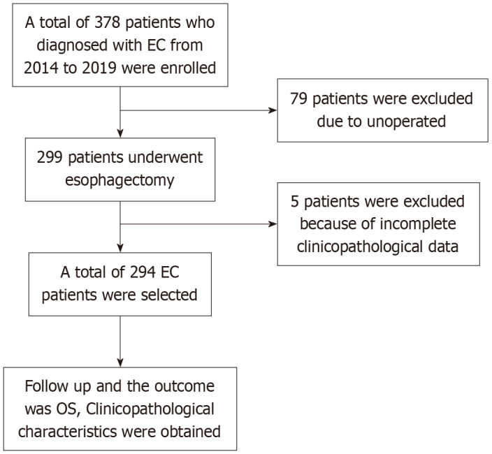
Methods: Serum lipid levels and clinicopathological information were retrospectively collected from 294 patients, and risk factors for LN metastasis were confirmed using a logistic regression model. Latent factors were explored using information from publicly accessible databases and immunofluorescence and immunohistochemical staining techniques.
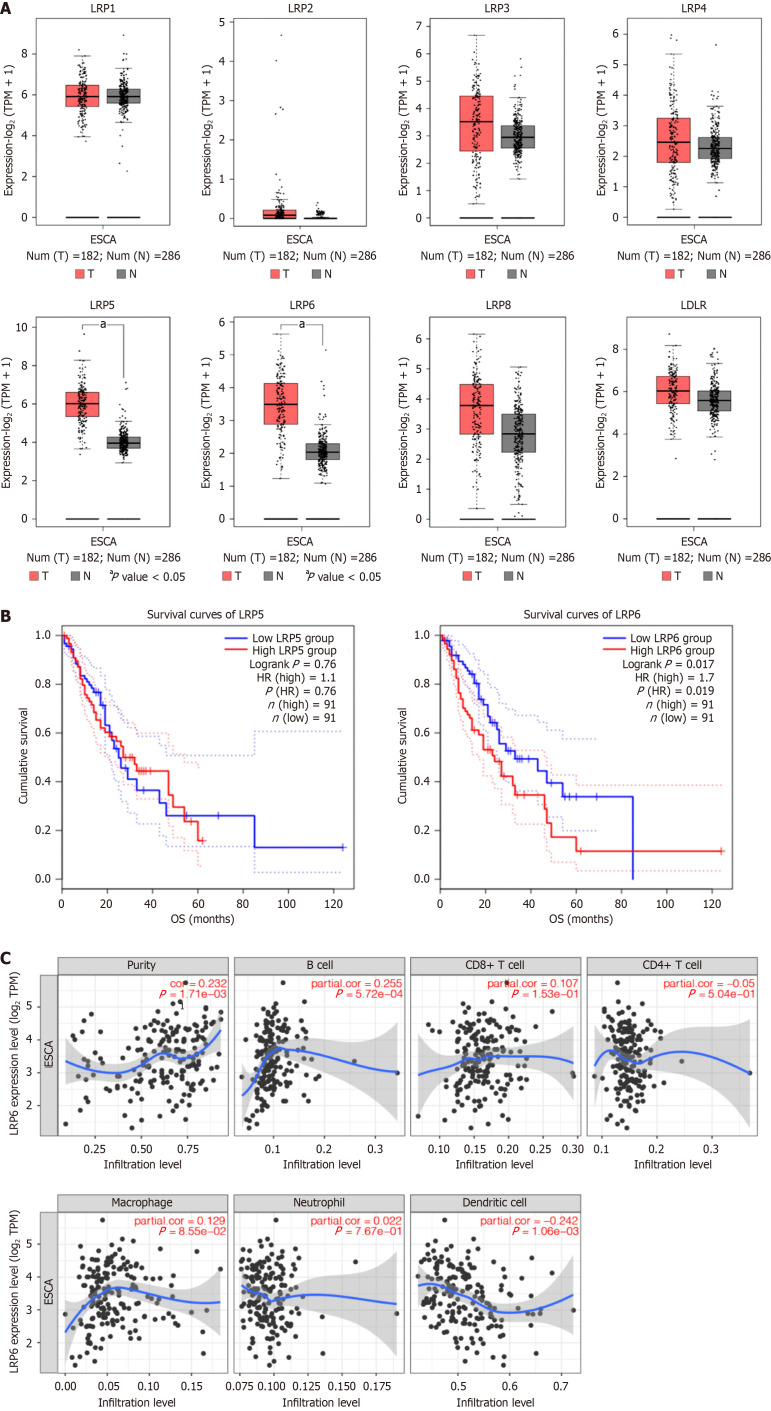
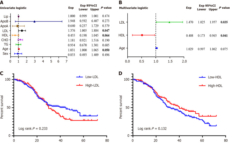
Results: High serum levels of low-density lipoprotein (LDL) cholesterol promote LN metastasis in EC, while high-density lipoprotein cholesterol has the opposite role. Information of a public database revealed that LDL receptors LRP5 and LRP6 are highly expressed in ECs, and LRP6 overexpression positively correlated with the infiltration of B lymphocytes and a poor prognosis. Immunofluorescence and immunohistochemical staining revealed that the expression of LRP6 and infiltrated B lymphocytes in patients with ≥ 1 regional LN metastasis, containing N1-3 (N+ group) were significantly higher than those in the N0 group. LRP6 was also highly expressed in the B lymphocytes of the N+ group. There was no difference in CXCL13 expression between the N+ and N0 groups. However, CXCR5 expression was significantly higher in the N0 group than in the N+ group.
Conclusion: High serum LDL levels can promote LN metastasis in EC, and the mechanisms may be related to LRP6 expression and the infiltration of B lymphocytes.

 求助内容:
求助内容: 应助结果提醒方式:


