Mohamad Ayham Muqresh, Sandrella I Zebian, Abdulkarim M Alkadrou, Osama Al-Shoaib, Thamer Bin Traiki
{"title":"Right Hepatic Lobe Hypoplasia and Gangrenous Cholecystitis: Surgical Implications and Case Report.","authors":"Mohamad Ayham Muqresh, Sandrella I Zebian, Abdulkarim M Alkadrou, Osama Al-Shoaib, Thamer Bin Traiki","doi":"10.12659/AJCR.948437","DOIUrl":null,"url":null,"abstract":"<p><p>BACKGROUND Hypoplasia of the right hepatic lobe is a rare congenital anomaly often discovered incidentally during imaging or surgery. This anomaly can be challenging during surgery, leading to intraoperative complications. Patient with right hepatic hypoplasia usually live normally without symptoms, but it can be associated with liver cirrhosis, portal hypertension, and gallstones. While the exact pathophysiological relationship between congenital hepatic hypoplasia and cholecystitis remains unclear, a few contributing factors have been proposed. These include mutations in the hepatocyte nuclear factor 1B (HNF1B) transcription, impaired gallbladder function, and gallbladder ischemia due to malposition. CASE REPORT A 70-year-old man presented with gangrenous cholecystitis and concurrent hypoplasia of the right hepatic lobe; a combination not previously documented. The patient, initially asymptomatic, presented with sharp epigastric pain and was diagnosed with acute cholecystitis through abdominal ultrasound. However, computed tomography (CT) imaging showed severe hypoplasia of the right liver lobe with associated compensatory hypertrophy of the left lobe in addition to stones in the gallbladder. Despite normal laboratory results, we think that the anomaly delayed the diagnosis until the development of cholecystitis as a complication. The patient underwent a successful laparoscopic cholecystectomy without converting to open surgery. CONCLUSIONS This case underscores the importance of recognizing anatomical variations when planning surgery. Awareness of right hepatic lobe hypoplasia can aid in timely diagnosis and appropriate management, ultimately reducing surgical risks and complications. Furthermore, we recommend performing CT in case of vague or atypical symptoms of acute cholecystitis, as well as when the gallbladder shows malposition on abdominal ultrasound (US). However, we recommend identifying the anatomical structure during the surgery to avoid complications.</p>","PeriodicalId":39064,"journal":{"name":"American Journal of Case Reports","volume":"26 ","pages":"e948437"},"PeriodicalIF":0.7000,"publicationDate":"2025-09-14","publicationTypes":"Journal Article","fieldsOfStudy":null,"isOpenAccess":false,"openAccessPdf":"https://www.ncbi.nlm.nih.gov/pmc/articles/PMC12445154/pdf/","citationCount":"0","resultStr":null,"platform":"Semanticscholar","paperid":null,"PeriodicalName":"American Journal of Case Reports","FirstCategoryId":"1085","ListUrlMain":"https://doi.org/10.12659/AJCR.948437","RegionNum":0,"RegionCategory":null,"ArticlePicture":[],"TitleCN":null,"AbstractTextCN":null,"PMCID":null,"EPubDate":"","PubModel":"","JCR":"Q3","JCRName":"MEDICINE, GENERAL & INTERNAL","Score":null,"Total":0}
引用次数: 0
Abstract
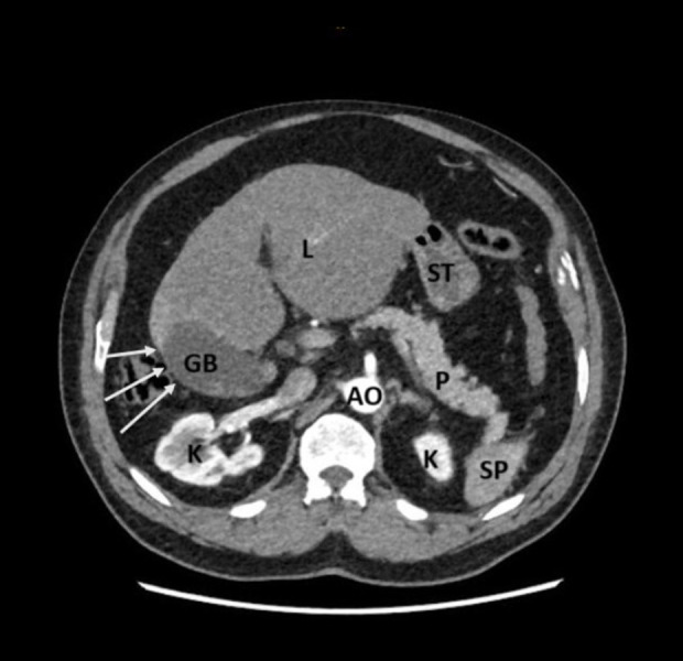
BACKGROUND Hypoplasia of the right hepatic lobe is a rare congenital anomaly often discovered incidentally during imaging or surgery. This anomaly can be challenging during surgery, leading to intraoperative complications. Patient with right hepatic hypoplasia usually live normally without symptoms, but it can be associated with liver cirrhosis, portal hypertension, and gallstones. While the exact pathophysiological relationship between congenital hepatic hypoplasia and cholecystitis remains unclear, a few contributing factors have been proposed. These include mutations in the hepatocyte nuclear factor 1B (HNF1B) transcription, impaired gallbladder function, and gallbladder ischemia due to malposition. CASE REPORT A 70-year-old man presented with gangrenous cholecystitis and concurrent hypoplasia of the right hepatic lobe; a combination not previously documented. The patient, initially asymptomatic, presented with sharp epigastric pain and was diagnosed with acute cholecystitis through abdominal ultrasound. However, computed tomography (CT) imaging showed severe hypoplasia of the right liver lobe with associated compensatory hypertrophy of the left lobe in addition to stones in the gallbladder. Despite normal laboratory results, we think that the anomaly delayed the diagnosis until the development of cholecystitis as a complication. The patient underwent a successful laparoscopic cholecystectomy without converting to open surgery. CONCLUSIONS This case underscores the importance of recognizing anatomical variations when planning surgery. Awareness of right hepatic lobe hypoplasia can aid in timely diagnosis and appropriate management, ultimately reducing surgical risks and complications. Furthermore, we recommend performing CT in case of vague or atypical symptoms of acute cholecystitis, as well as when the gallbladder shows malposition on abdominal ultrasound (US). However, we recommend identifying the anatomical structure during the surgery to avoid complications.
期刊介绍:
American Journal of Case Reports is an international, peer-reviewed scientific journal that publishes single and series case reports in all medical fields. American Journal of Case Reports is issued on a continuous basis as a primary electronic journal. Print copies of a single article or a set of articles can be ordered on demand.

 求助内容:
求助内容: 应助结果提醒方式:


