Rafael Netto, Maria Elisa Rangel Janini, Wladimir Cortezzi, Ludimila Lemes Moura, Silas Antonio Juvencio de Freitas-Filho
{"title":"Keratoameloblastoma of the jaws and review of international literature of 38 cases.","authors":"Rafael Netto, Maria Elisa Rangel Janini, Wladimir Cortezzi, Ludimila Lemes Moura, Silas Antonio Juvencio de Freitas-Filho","doi":"10.4317/jced.62973","DOIUrl":null,"url":null,"abstract":"<p><p>The term keratoameloblastoma has been used to describe a histologically heterogeneous group of ameloblastoma variants that share the formation of keratin by the ameloblastomatous epithelium. To date, thirty-eight cases of keratoameloblastoma have been previously reported in the literature, nine of which exhibited a papilliferous component. Here we report a new case of a recurrent tumor that falls within the keratoameloblastoma spectrum. It presented as an expansile, solid lesion with internal calcification in the right infratemporal fossa six years after ipsilateral hemimandibulectomy in a 46-year-old white female. Histological evaluation revealed islands of columnar cells resembling ameloblasts surrounding a central area with stellate reticulum-like cells, some of them completely filled with keratin. In addition, areas showed basal ranging from columnar to cuboidal with hyperchromatic nuclei. The clinical, histopathologic, and radiographic features of keratoameloblastoma are reviewed, along with treatment approaches and follow-up considerations. Although only a few cases have been documented, the tumor's aggressive biological behavior and the high recurrence rate suggest that a more aggressive therapeutic approach is warranted. Patients should be informed of the importance of clinical monitoring. Surgical resection with adequate safety margins and histopathological evaluation of the margins is strongly recommended. <b>Key words:</b>Odontogenic tumors, keratoameloblastoma, ameloblastoma, review.</p>","PeriodicalId":15376,"journal":{"name":"Journal of Clinical and Experimental Dentistry","volume":"17 8","pages":"e1006-e1013"},"PeriodicalIF":0.0000,"publicationDate":"2025-08-01","publicationTypes":"Journal Article","fieldsOfStudy":null,"isOpenAccess":false,"openAccessPdf":"https://www.ncbi.nlm.nih.gov/pmc/articles/PMC12424603/pdf/","citationCount":"0","resultStr":null,"platform":"Semanticscholar","paperid":null,"PeriodicalName":"Journal of Clinical and Experimental Dentistry","FirstCategoryId":"1085","ListUrlMain":"https://doi.org/10.4317/jced.62973","RegionNum":0,"RegionCategory":null,"ArticlePicture":[],"TitleCN":null,"AbstractTextCN":null,"PMCID":null,"EPubDate":"","PubModel":"","JCR":"Q2","JCRName":"Dentistry","Score":null,"Total":0}
引用次数: 0
Abstract
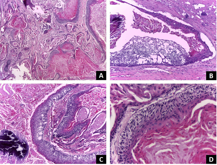
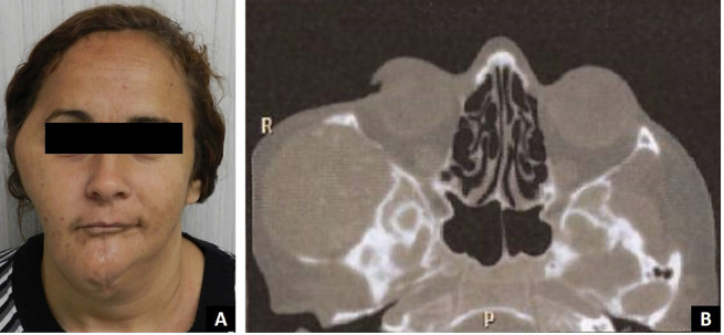
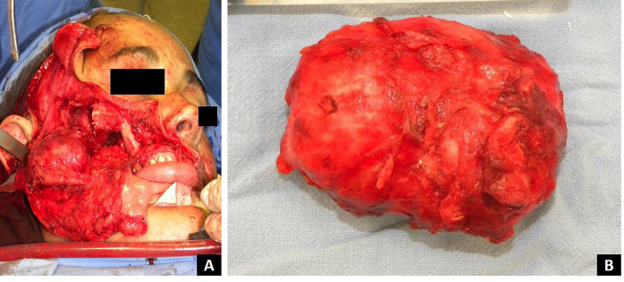
The term keratoameloblastoma has been used to describe a histologically heterogeneous group of ameloblastoma variants that share the formation of keratin by the ameloblastomatous epithelium. To date, thirty-eight cases of keratoameloblastoma have been previously reported in the literature, nine of which exhibited a papilliferous component. Here we report a new case of a recurrent tumor that falls within the keratoameloblastoma spectrum. It presented as an expansile, solid lesion with internal calcification in the right infratemporal fossa six years after ipsilateral hemimandibulectomy in a 46-year-old white female. Histological evaluation revealed islands of columnar cells resembling ameloblasts surrounding a central area with stellate reticulum-like cells, some of them completely filled with keratin. In addition, areas showed basal ranging from columnar to cuboidal with hyperchromatic nuclei. The clinical, histopathologic, and radiographic features of keratoameloblastoma are reviewed, along with treatment approaches and follow-up considerations. Although only a few cases have been documented, the tumor's aggressive biological behavior and the high recurrence rate suggest that a more aggressive therapeutic approach is warranted. Patients should be informed of the importance of clinical monitoring. Surgical resection with adequate safety margins and histopathological evaluation of the margins is strongly recommended. Key words:Odontogenic tumors, keratoameloblastoma, ameloblastoma, review.
期刊介绍:
Indexed in PUBMED, PubMed Central® (PMC) since 2012 and SCOPUSJournal of Clinical and Experimental Dentistry is an Open Access (free access on-line) - http://www.medicinaoral.com/odo/indice.htm. The aim of the Journal of Clinical and Experimental Dentistry is: - Periodontology - Community and Preventive Dentistry - Esthetic Dentistry - Biomaterials and Bioengineering in Dentistry - Operative Dentistry and Endodontics - Prosthetic Dentistry - Orthodontics - Oral Medicine and Pathology - Odontostomatology for the disabled or special patients - Oral Surgery

 求助内容:
求助内容: 应助结果提醒方式:


