AmirHossein SohrabiFar, Donya Maleki, Arayeh Maleki, Helia Zare, Dina Maleki
{"title":"The Relation of Skeletal Malocclusion and Airway Volumes: A Cross-Sectional Study.","authors":"AmirHossein SohrabiFar, Donya Maleki, Arayeh Maleki, Helia Zare, Dina Maleki","doi":"10.1155/ijod/2318588","DOIUrl":null,"url":null,"abstract":"<p><p><b>Aim:</b> This study evaluated the relationship between airway volume and skeletal malocclusion. <b>Methods:</b> This study was a cross-sectional analytical study obtaining 450 cone-beam computed tomography (CBCT) images from the archives of a private clinic taken by the Sirona Galileos Comfort Plus, Dentsply Sirona, Germany device, following the Fast-Scan protocol with 14 s exposure time, FOV = 15 × 15 cm, kV = 98, and mA = 3. The CBCT images were from adults aged 17-39 years with a normal pattern in their vertical growth (SN-GO.GN = 32 ± 5), with no history of orthognathic or rhino surgery, no syndromes, no previous trauma, and no pathologies along the airway and pharynx detectable in the images. CBCT images with radiographical artifacts and low quality or resolution were excluded from the study. The total pharyngeal volume (TPV) was measured from the superior part of the PNS (posterior nasal spine) parallel to the standard horizontal plane to the anterior-inferior part of the C4 vertebra, parallel to the standard horizontal plane. Velopharynx volume (VPV) was measured from the superior part of the PNS to the inferior border of the soft palate. Glossopharynx volume (GPV) was measured from the inferior border of the soft palate to the superior tip of the epiglottis. The volumes were reported in mm<sup>3</sup>. To analyze data, SPSS version 22.0 (IBM Corp, Armonk, NY, USA) was used. ANOVA and Tukey's post hoc test were applied. <b>Results:</b> The results revealed that the mean total pharyngeal airway volume and velopharyngeal airway volume were significantly larger in Class III patients compared to Class II and Class I patients. Also, in Class I patients, the mean total pharyngeal airway volume and velopharyngeal airway volume were significantly greater than in Class II patients. The glossopharynx airway volume was significantly different between Class II and Class III patients, so the glossopharynx airway volume was significantly greater in Class III patients than in Class II patients. <b>Conclusion:</b> The results showed that there is a relationship between skeletal malocclusion and airway volumes.</p>","PeriodicalId":13947,"journal":{"name":"International Journal of Dentistry","volume":"2025 ","pages":"2318588"},"PeriodicalIF":2.2000,"publicationDate":"2025-09-05","publicationTypes":"Journal Article","fieldsOfStudy":null,"isOpenAccess":false,"openAccessPdf":"https://www.ncbi.nlm.nih.gov/pmc/articles/PMC12431819/pdf/","citationCount":"0","resultStr":null,"platform":"Semanticscholar","paperid":null,"PeriodicalName":"International Journal of Dentistry","FirstCategoryId":"1085","ListUrlMain":"https://doi.org/10.1155/ijod/2318588","RegionNum":0,"RegionCategory":null,"ArticlePicture":[],"TitleCN":null,"AbstractTextCN":null,"PMCID":null,"EPubDate":"2025/1/1 0:00:00","PubModel":"eCollection","JCR":"Q2","JCRName":"DENTISTRY, ORAL SURGERY & MEDICINE","Score":null,"Total":0}
引用次数: 0
Abstract
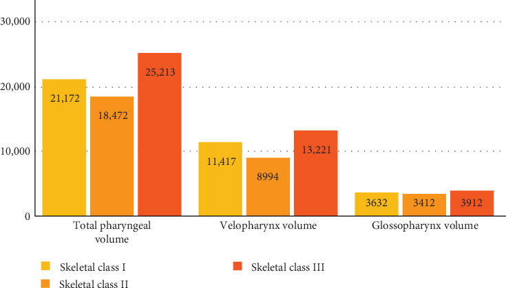
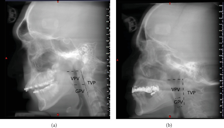
Aim: This study evaluated the relationship between airway volume and skeletal malocclusion. Methods: This study was a cross-sectional analytical study obtaining 450 cone-beam computed tomography (CBCT) images from the archives of a private clinic taken by the Sirona Galileos Comfort Plus, Dentsply Sirona, Germany device, following the Fast-Scan protocol with 14 s exposure time, FOV = 15 × 15 cm, kV = 98, and mA = 3. The CBCT images were from adults aged 17-39 years with a normal pattern in their vertical growth (SN-GO.GN = 32 ± 5), with no history of orthognathic or rhino surgery, no syndromes, no previous trauma, and no pathologies along the airway and pharynx detectable in the images. CBCT images with radiographical artifacts and low quality or resolution were excluded from the study. The total pharyngeal volume (TPV) was measured from the superior part of the PNS (posterior nasal spine) parallel to the standard horizontal plane to the anterior-inferior part of the C4 vertebra, parallel to the standard horizontal plane. Velopharynx volume (VPV) was measured from the superior part of the PNS to the inferior border of the soft palate. Glossopharynx volume (GPV) was measured from the inferior border of the soft palate to the superior tip of the epiglottis. The volumes were reported in mm3. To analyze data, SPSS version 22.0 (IBM Corp, Armonk, NY, USA) was used. ANOVA and Tukey's post hoc test were applied. Results: The results revealed that the mean total pharyngeal airway volume and velopharyngeal airway volume were significantly larger in Class III patients compared to Class II and Class I patients. Also, in Class I patients, the mean total pharyngeal airway volume and velopharyngeal airway volume were significantly greater than in Class II patients. The glossopharynx airway volume was significantly different between Class II and Class III patients, so the glossopharynx airway volume was significantly greater in Class III patients than in Class II patients. Conclusion: The results showed that there is a relationship between skeletal malocclusion and airway volumes.

 求助内容:
求助内容: 应助结果提醒方式:


