Chiara Sordelli, Carlo Liguori, Nunzia Fele, Sara Hana Weisz, Raffaele Verde, Angela Guarino, Nunzia De Crescenzo, Alessandro Perrella, Emilio Di Lorenzo
{"title":"Perivalvular Complication in Infective Endocarditis: An Integrated Imaging Approach in the Diagnostic Workup.","authors":"Chiara Sordelli, Carlo Liguori, Nunzia Fele, Sara Hana Weisz, Raffaele Verde, Angela Guarino, Nunzia De Crescenzo, Alessandro Perrella, Emilio Di Lorenzo","doi":"10.4103/jcecho.jcecho_92_24","DOIUrl":null,"url":null,"abstract":"<p><p>Infective endocarditis (IE) is a rare disease, but its impact is significant as it affects 3-10/100,000 per year in the population. According to the current guidelines ESC 2023, the evidence of lesions characteristic of IE is a major diagnostic criterion. Echocardiography is the first-line imaging technique to diagnose IE and to assess the structural and functional damage of cardiac structures. Transesophageal echocardiography (TEE) is recommended in patients with an inconclusive or negative TTE, in patients with high suspicion of IE, as well as in patients with a positive TTE, to document local complications. Other imaging modalities, such as cardiac computed tomography and nuclear imaging, are needed to confirm or exclude the diagnosis of IE, to characterize the extent of the cardiac lesions, and to diagnose cardiac complications. The aim of this article was to review the potential role of cardiac imaging, especially of TEE and cardiac CT in evaluating IE perivalvular complications.</p>","PeriodicalId":15191,"journal":{"name":"Journal of Cardiovascular Echography","volume":"35 2","pages":"108-115"},"PeriodicalIF":1.0000,"publicationDate":"2025-04-01","publicationTypes":"Journal Article","fieldsOfStudy":null,"isOpenAccess":false,"openAccessPdf":"https://www.ncbi.nlm.nih.gov/pmc/articles/PMC12425274/pdf/","citationCount":"0","resultStr":null,"platform":"Semanticscholar","paperid":null,"PeriodicalName":"Journal of Cardiovascular Echography","FirstCategoryId":"1085","ListUrlMain":"https://doi.org/10.4103/jcecho.jcecho_92_24","RegionNum":0,"RegionCategory":null,"ArticlePicture":[],"TitleCN":null,"AbstractTextCN":null,"PMCID":null,"EPubDate":"2025/7/30 0:00:00","PubModel":"Epub","JCR":"Q4","JCRName":"CARDIAC & CARDIOVASCULAR SYSTEMS","Score":null,"Total":0}
引用次数: 0
Abstract
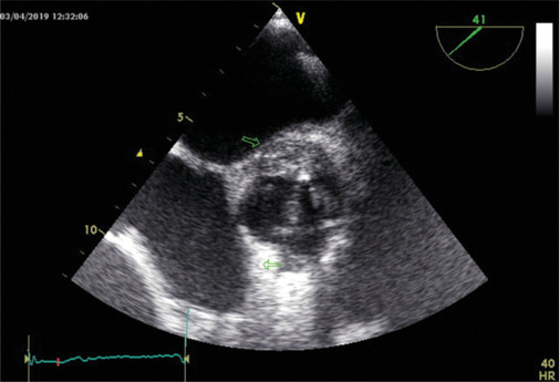
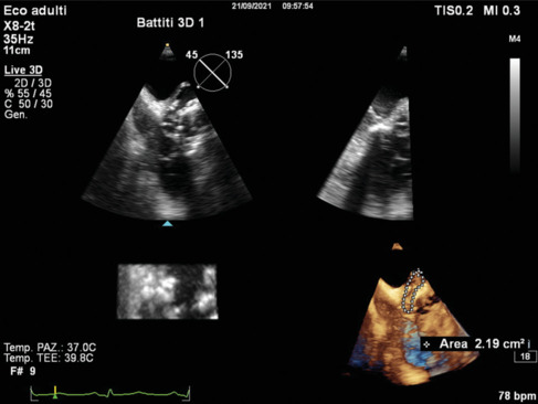
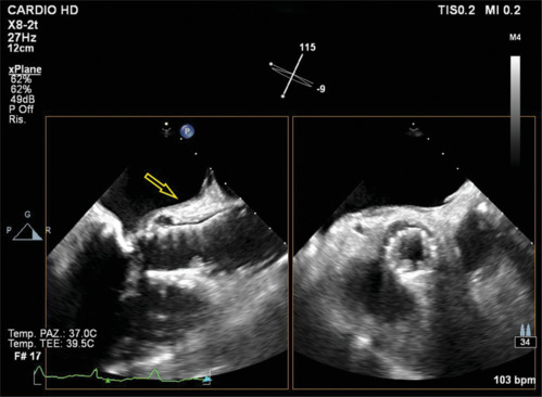
Infective endocarditis (IE) is a rare disease, but its impact is significant as it affects 3-10/100,000 per year in the population. According to the current guidelines ESC 2023, the evidence of lesions characteristic of IE is a major diagnostic criterion. Echocardiography is the first-line imaging technique to diagnose IE and to assess the structural and functional damage of cardiac structures. Transesophageal echocardiography (TEE) is recommended in patients with an inconclusive or negative TTE, in patients with high suspicion of IE, as well as in patients with a positive TTE, to document local complications. Other imaging modalities, such as cardiac computed tomography and nuclear imaging, are needed to confirm or exclude the diagnosis of IE, to characterize the extent of the cardiac lesions, and to diagnose cardiac complications. The aim of this article was to review the potential role of cardiac imaging, especially of TEE and cardiac CT in evaluating IE perivalvular complications.

 求助内容:
求助内容: 应助结果提醒方式:


