{"title":"Septo-Optic Dysplasia Plus with Bilateral Homonymous Hemianopia - Case Report.","authors":"Jan Lestak, Martin Fus, Martin Kyncl","doi":"10.2147/IMCRJ.S513192","DOIUrl":null,"url":null,"abstract":"<p><p>The paper presents a case report of a man (born1968) who was examined for unusual changes in the visual fields. During the examination in 2020, the right eye had esotropia, the pupillary reaction was correct. The ocular findings on the anterior segment and the ocular media were normal. The papilla of the right eye was temporally paler with c/d=0.5, the left papilla was pale with c/d=0.6, otherwise the background was normal. IOP: 16/18 mmHg. During the examination, diffuse loss of the nerve fiber layer (50 um, resp. 49 um) was detected using OCT, and similarly low values were also found for vessel density. The visual fields showed left-sided homonymous hemianopia with central sparing, partially extending into the upper right quadrants. Electrophysiological examinations of the retina (pattern electroretinogram) and the entire visual analyzer (pattern visual evoked potential - PVEP) showed bilaterally lower amplitudes. The latencies of the P00 VEP peak were not prolonged. For these changes, a magnetic resonance imaging (MRI) examination of the brain was performed with the finding of agenesis of the corpus callosum, associated trigone of the lateral ventricles on both sides. Malformation of the development of the cerebral cortex temporooccipitally medially on the right, of the nature of plymicrogyria. Heterotopia of gray matter periventricularly occipitally on the right. Bilateral atrophy of the optic nerves and chiasm. In the case report of SOD plus, unusual changes in the visual fields are described - homonymous left-sided hemianopia with central sparing. MRI examination of the brain helped to classify this lesion in the temporo-occipital medial region on the right with polymicrogyria.</p>","PeriodicalId":14337,"journal":{"name":"International Medical Case Reports Journal","volume":"18 ","pages":"1159-1165"},"PeriodicalIF":0.7000,"publicationDate":"2025-09-08","publicationTypes":"Journal Article","fieldsOfStudy":null,"isOpenAccess":false,"openAccessPdf":"https://www.ncbi.nlm.nih.gov/pmc/articles/PMC12430242/pdf/","citationCount":"0","resultStr":null,"platform":"Semanticscholar","paperid":null,"PeriodicalName":"International Medical Case Reports Journal","FirstCategoryId":"1085","ListUrlMain":"https://doi.org/10.2147/IMCRJ.S513192","RegionNum":0,"RegionCategory":null,"ArticlePicture":[],"TitleCN":null,"AbstractTextCN":null,"PMCID":null,"EPubDate":"2025/1/1 0:00:00","PubModel":"eCollection","JCR":"Q3","JCRName":"MEDICINE, GENERAL & INTERNAL","Score":null,"Total":0}
引用次数: 0
Abstract
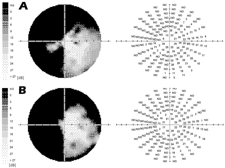
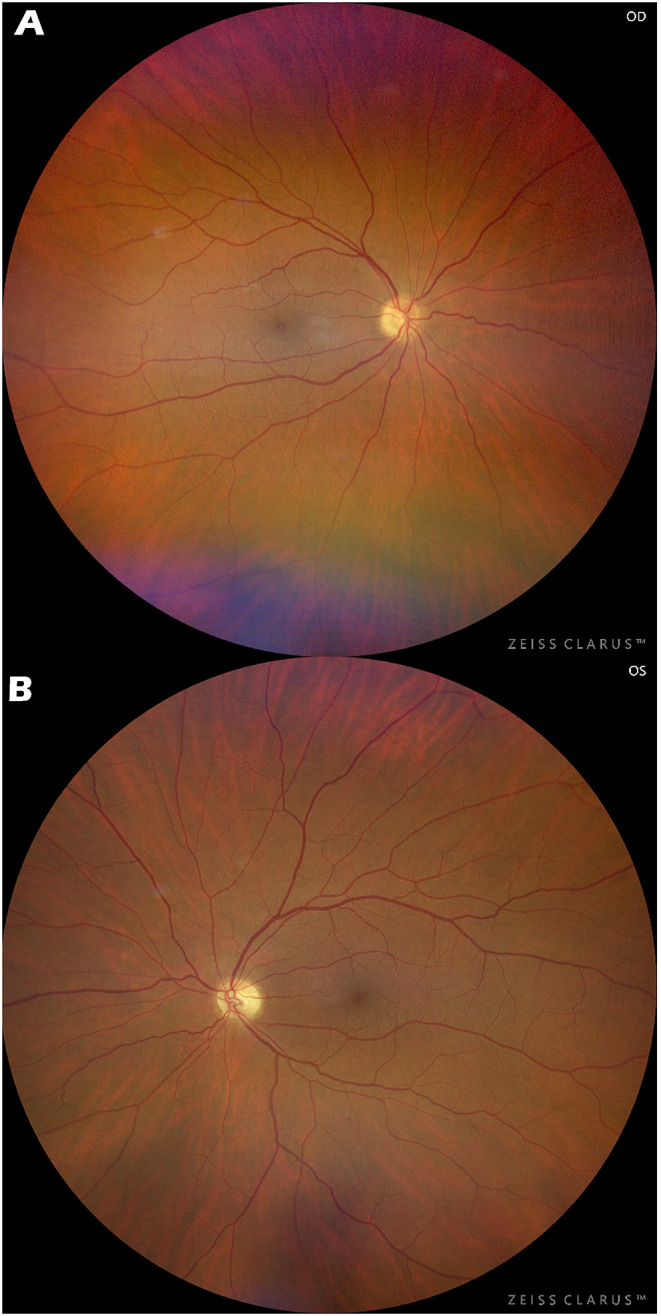
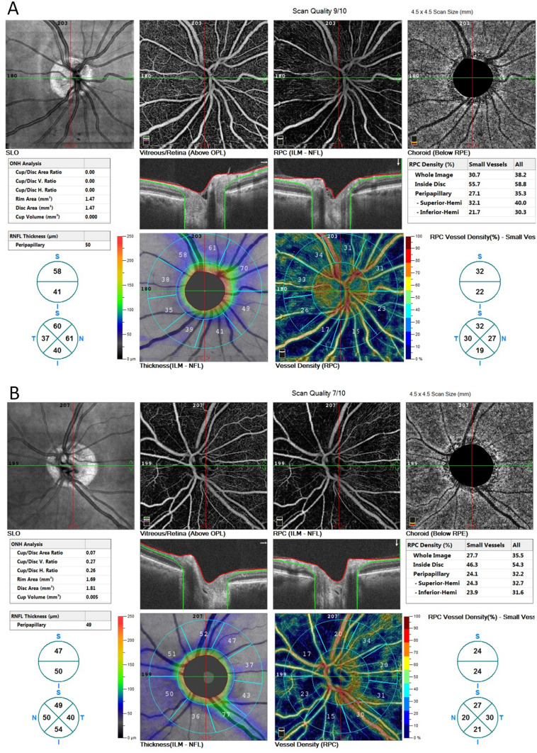
The paper presents a case report of a man (born1968) who was examined for unusual changes in the visual fields. During the examination in 2020, the right eye had esotropia, the pupillary reaction was correct. The ocular findings on the anterior segment and the ocular media were normal. The papilla of the right eye was temporally paler with c/d=0.5, the left papilla was pale with c/d=0.6, otherwise the background was normal. IOP: 16/18 mmHg. During the examination, diffuse loss of the nerve fiber layer (50 um, resp. 49 um) was detected using OCT, and similarly low values were also found for vessel density. The visual fields showed left-sided homonymous hemianopia with central sparing, partially extending into the upper right quadrants. Electrophysiological examinations of the retina (pattern electroretinogram) and the entire visual analyzer (pattern visual evoked potential - PVEP) showed bilaterally lower amplitudes. The latencies of the P00 VEP peak were not prolonged. For these changes, a magnetic resonance imaging (MRI) examination of the brain was performed with the finding of agenesis of the corpus callosum, associated trigone of the lateral ventricles on both sides. Malformation of the development of the cerebral cortex temporooccipitally medially on the right, of the nature of plymicrogyria. Heterotopia of gray matter periventricularly occipitally on the right. Bilateral atrophy of the optic nerves and chiasm. In the case report of SOD plus, unusual changes in the visual fields are described - homonymous left-sided hemianopia with central sparing. MRI examination of the brain helped to classify this lesion in the temporo-occipital medial region on the right with polymicrogyria.
期刊介绍:
International Medical Case Reports Journal is an international, peer-reviewed, open access, online journal publishing original case reports from all medical specialties. Submissions should not normally exceed 3,000 words or 4 published pages including figures, diagrams and references. As of 1st April 2019, the International Medical Case Reports Journal will no longer consider meta-analyses for publication.

 求助内容:
求助内容: 应助结果提醒方式:


