Thamil Selvan Muthuraj, Renganath Murugan Jeyasree, Gokulvathi Rajkumar, Jacob Raja, Johnson Raja James, Jeyaraj Prince Mohanraj
{"title":"Comparative evaluation of ultrasonography with cone-beam computed tomography for peri-implant bone loss analysis - A cadaveric goat mandible study.","authors":"Thamil Selvan Muthuraj, Renganath Murugan Jeyasree, Gokulvathi Rajkumar, Jacob Raja, Johnson Raja James, Jeyaraj Prince Mohanraj","doi":"10.4103/jisp.jisp_83_24","DOIUrl":null,"url":null,"abstract":"<p><strong>Background: </strong>Early diagnosis of peri-implant bone loss is vital for the successful management of peri-implantitis. Among the various diagnostic tools available, cone-beam computed tomography (CBCT) is the most commonly used method. However, CBCT has own limitations, which have led to the development of new diagnostic technologies. Of these, ultrasonography (US) appears to be the most promising. The aim of the current study was to compare and evaluate the efficacy of US with CBCT and direct measurement (DM) using a periodontal probe (PP) in assessing peri-implant bone defects created in a cadaveric goat mandible.</p><p><strong>Materials and methods: </strong>Fifteen cadaveric goat mandibles were procured from a local slaughterhouse, and 15 endosseous implants (3.5 mm × 13 mm) were placed using a standard surgical protocol. Critical defects measuring 7 mm × 3 mm × 3 mm were created around the implants using a piezosurgical unit. These defects were evaluated using a PP for DM in Group I (GI), CBCT in Group II (GII), and ultrasonography (US) in Group III (GIII). The collected data were statistically analyzed.</p><p><strong>Results: </strong>The mean height of the peri-implant osseous defect measured in GI, GII, and GIII was 6.05 ± 0.08 mm, 5.55 ± 0.40 mm, and 6.05 ± 0.30 mm, respectively. The measurements in GIII were comparable to those of GI and more precise than those of GII.</p><p><strong>Conclusion: </strong>Within the limitations of the current study, US is more effective than CBCT and equally effective as DM using a PP in measuring critical peri-implant bone defects in cadaveric goat mandible. Therefore, ultrasonography can be considered a reliable diagnostic tool for routine implant follow-up.</p>","PeriodicalId":15890,"journal":{"name":"Journal of Indian Society of Periodontology","volume":"29 2","pages":"130-135"},"PeriodicalIF":0.0000,"publicationDate":"2025-03-01","publicationTypes":"Journal Article","fieldsOfStudy":null,"isOpenAccess":false,"openAccessPdf":"https://www.ncbi.nlm.nih.gov/pmc/articles/PMC12425252/pdf/","citationCount":"0","resultStr":null,"platform":"Semanticscholar","paperid":null,"PeriodicalName":"Journal of Indian Society of Periodontology","FirstCategoryId":"1085","ListUrlMain":"https://doi.org/10.4103/jisp.jisp_83_24","RegionNum":0,"RegionCategory":null,"ArticlePicture":[],"TitleCN":null,"AbstractTextCN":null,"PMCID":null,"EPubDate":"2025/8/19 0:00:00","PubModel":"Epub","JCR":"Q2","JCRName":"Dentistry","Score":null,"Total":0}
引用次数: 0
Abstract
Background: Early diagnosis of peri-implant bone loss is vital for the successful management of peri-implantitis. Among the various diagnostic tools available, cone-beam computed tomography (CBCT) is the most commonly used method. However, CBCT has own limitations, which have led to the development of new diagnostic technologies. Of these, ultrasonography (US) appears to be the most promising. The aim of the current study was to compare and evaluate the efficacy of US with CBCT and direct measurement (DM) using a periodontal probe (PP) in assessing peri-implant bone defects created in a cadaveric goat mandible.
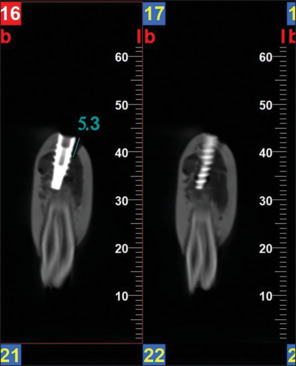
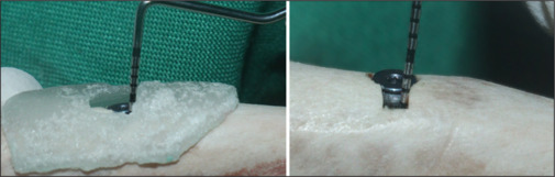
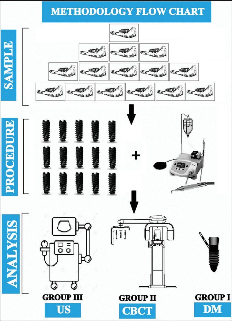
Materials and methods: Fifteen cadaveric goat mandibles were procured from a local slaughterhouse, and 15 endosseous implants (3.5 mm × 13 mm) were placed using a standard surgical protocol. Critical defects measuring 7 mm × 3 mm × 3 mm were created around the implants using a piezosurgical unit. These defects were evaluated using a PP for DM in Group I (GI), CBCT in Group II (GII), and ultrasonography (US) in Group III (GIII). The collected data were statistically analyzed.
Results: The mean height of the peri-implant osseous defect measured in GI, GII, and GIII was 6.05 ± 0.08 mm, 5.55 ± 0.40 mm, and 6.05 ± 0.30 mm, respectively. The measurements in GIII were comparable to those of GI and more precise than those of GII.
Conclusion: Within the limitations of the current study, US is more effective than CBCT and equally effective as DM using a PP in measuring critical peri-implant bone defects in cadaveric goat mandible. Therefore, ultrasonography can be considered a reliable diagnostic tool for routine implant follow-up.
期刊介绍:
The Journal of Indian Society of Periodontology publishes original scientific articles to support practice , education and research in the dental specialty of periodontology and oral implantology. Journal of Indian Society of Periodontology (JISP), is the official publication of the Society and is managed and brought out by the Editor of the society. The journal is published Bimonthly with special issues being brought out for specific occasions. The ISP had a bulletin as its publication for a large number of years and was enhanced as a Journal a few years ago

 求助内容:
求助内容: 应助结果提醒方式:


