Salvatore Gitto, Anna Corti, Kirsten van Langevelde, Ana Navas Cañete, Antonino Cincotta, Carmelo Messina, Domenico Albano, Carlotta Vignaga, Laura Ferrari, Luca Mainardi, Valentina D A Corino, Luca Maria Sconfienza
{"title":"Deep learning for automated segmentation of central cartilage tumors on MRI.","authors":"Salvatore Gitto, Anna Corti, Kirsten van Langevelde, Ana Navas Cañete, Antonino Cincotta, Carmelo Messina, Domenico Albano, Carlotta Vignaga, Laura Ferrari, Luca Mainardi, Valentina D A Corino, Luca Maria Sconfienza","doi":"10.1186/s41747-025-00633-7","DOIUrl":null,"url":null,"abstract":"<p><strong>Background: </strong>Automated segmentation methods may potentially increase the reliability and applicability of radiomics in skeletal oncology. Our aim was to propose a deep learning-based method for automated segmentation of atypical cartilaginous tumor (ACT) and grade II chondrosarcoma (CS2) of long bones on magnetic resonance imaging (MRI).</p><p><strong>Materials and methods: </strong>This institutional review board-approved retrospective study included 164 patients with surgically treated and histology-proven cartilaginous tumors at two tertiary bone tumor centers. The first cohort consisted of 99 MRI scans from center 1 (79 ACT, 20 CS2). The second cohort consisted of 65 MRI scans from center 2 (45 ACT, 20 CS2). Supervised Edge-Attention Guidance segmentation Network (SEAGNET) architecture was employed for automated image segmentation on T1-weighted images, using manual segmentations drawn by musculoskeletal radiologists as the ground truth. In the first cohort, a total of 1,037 slices containing the tumor out of 99 patients were split into 70% training, 15% validation, and 15% internal test sets, respectively, and used for model tuning. The second cohort was used for independent external testing.</p><p><strong>Results: </strong>In the first cohort, Dice Score (DS) and Intersection over Union (IoU) per patient were 0.782 ± 0.148 and 0.663 ± 0.175, and 0.748 ± 0.191 and 0.630 ± 0.210 in the validation and internal test sets, respectively. DS and IoU per slice were 0.742 ± 0.273 and 0.646 ± 0.266, and 0.752 ± 0.256 and 0.656 ± 0.261 in the validation and internal test sets, respectively. In the independent external test dataset, the model achieved DS of 0.828 ± 0.175 and IoU of 0.706 ± 0.180.</p><p><strong>Conclusion: </strong>Deep learning proved excellent for automated segmentation of central cartilage tumors on MRI.</p><p><strong>Relevance statement: </strong>A deep learning model based on SEAGNET architecture achieved excellent performance for automated segmentation of cartilage tumors of long bones on MRI and may be beneficial, given the increasing detection rate of these lesions in clinical practice.</p><p><strong>Key points: </strong>Automated segmentation may potentially increase the reliability and applicability of radiomics-based models. A deep learning architecture was proposed for automated segmentation of appendicular cartilage tumors on MRI. Deep learning proved excellent with a mean Dice Score of 0.828 in the external test cohort.</p>","PeriodicalId":36926,"journal":{"name":"European Radiology Experimental","volume":"9 1","pages":"91"},"PeriodicalIF":3.6000,"publicationDate":"2025-09-12","publicationTypes":"Journal Article","fieldsOfStudy":null,"isOpenAccess":false,"openAccessPdf":"https://www.ncbi.nlm.nih.gov/pmc/articles/PMC12431992/pdf/","citationCount":"0","resultStr":null,"platform":"Semanticscholar","paperid":null,"PeriodicalName":"European Radiology Experimental","FirstCategoryId":"1085","ListUrlMain":"https://doi.org/10.1186/s41747-025-00633-7","RegionNum":0,"RegionCategory":null,"ArticlePicture":[],"TitleCN":null,"AbstractTextCN":null,"PMCID":null,"EPubDate":"","PubModel":"","JCR":"Q1","JCRName":"RADIOLOGY, NUCLEAR MEDICINE & MEDICAL IMAGING","Score":null,"Total":0}
引用次数: 0
Abstract
Background: Automated segmentation methods may potentially increase the reliability and applicability of radiomics in skeletal oncology. Our aim was to propose a deep learning-based method for automated segmentation of atypical cartilaginous tumor (ACT) and grade II chondrosarcoma (CS2) of long bones on magnetic resonance imaging (MRI).
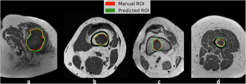
Materials and methods: This institutional review board-approved retrospective study included 164 patients with surgically treated and histology-proven cartilaginous tumors at two tertiary bone tumor centers. The first cohort consisted of 99 MRI scans from center 1 (79 ACT, 20 CS2). The second cohort consisted of 65 MRI scans from center 2 (45 ACT, 20 CS2). Supervised Edge-Attention Guidance segmentation Network (SEAGNET) architecture was employed for automated image segmentation on T1-weighted images, using manual segmentations drawn by musculoskeletal radiologists as the ground truth. In the first cohort, a total of 1,037 slices containing the tumor out of 99 patients were split into 70% training, 15% validation, and 15% internal test sets, respectively, and used for model tuning. The second cohort was used for independent external testing.
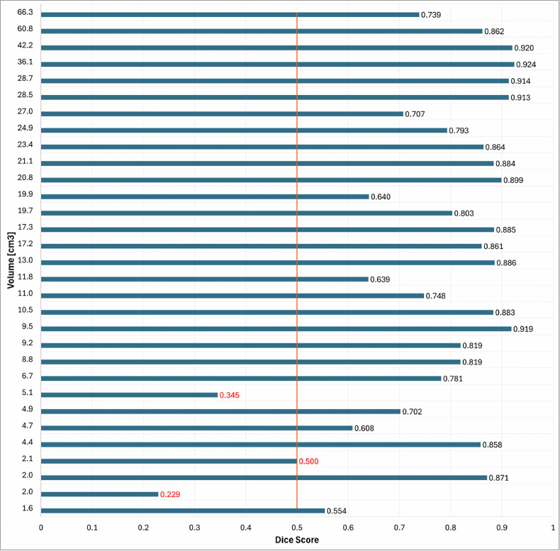
Results: In the first cohort, Dice Score (DS) and Intersection over Union (IoU) per patient were 0.782 ± 0.148 and 0.663 ± 0.175, and 0.748 ± 0.191 and 0.630 ± 0.210 in the validation and internal test sets, respectively. DS and IoU per slice were 0.742 ± 0.273 and 0.646 ± 0.266, and 0.752 ± 0.256 and 0.656 ± 0.261 in the validation and internal test sets, respectively. In the independent external test dataset, the model achieved DS of 0.828 ± 0.175 and IoU of 0.706 ± 0.180.
Conclusion: Deep learning proved excellent for automated segmentation of central cartilage tumors on MRI.
Relevance statement: A deep learning model based on SEAGNET architecture achieved excellent performance for automated segmentation of cartilage tumors of long bones on MRI and may be beneficial, given the increasing detection rate of these lesions in clinical practice.
Key points: Automated segmentation may potentially increase the reliability and applicability of radiomics-based models. A deep learning architecture was proposed for automated segmentation of appendicular cartilage tumors on MRI. Deep learning proved excellent with a mean Dice Score of 0.828 in the external test cohort.

 求助内容:
求助内容: 应助结果提醒方式:


