{"title":"Investigation of microstructural symmetry in regional zones of human multi-rooted teeth using optical, electrical, and ion diffusion methods.","authors":"Vladimir Mikhailovich Zolotarev","doi":"10.14440/jbm.2024.0122","DOIUrl":null,"url":null,"abstract":"<p><strong>Background: </strong>Dentin is a mineralized tissue characterized by a complex network of dentinal tubules, whose arrangement significantly influences the mechanical and physiological properties of teeth.</p><p><strong>Objective: </strong>This study investigated the influence of the microstructural symmetry of dentinal tubules in two orthogonal sections of the crown of human molar and premolar teeth.</p><p><strong>Methods: </strong>The effect of symmetry on the microstructure of dentin sections was studied for two orthogonal sections of the human molar and premolar crown. The symmetry of local zones of tooth sections was first examined using a set of methods: optical, electrical, and ion-diffusion techniques. The methods used have different resolutions and display both the general properties of the dentin structure and the properties that are specifically revealed by an individual method. It is shown that dentinal tubules originate from the center of the cusps of both molars and premolars, forming S-shaped fiber bundles presenting an axial-radial symmetry.</p><p><strong>Results: </strong>The dentinal tubules were shown to originate from the center of the cusps in both molar and premolar teeth, forming S-shaped fiber bundles with axial-radial symmetry. These bundles were arranged along axes, extending from the pulp toward the centers of the cusps of the tooth crown. Within these zones, distinct optical patterns resembling conoscopic figures in the form of a \"Maltese cross\" were observed. This indicates a highly ordered architecture composed of optically anisotropic uniaxial tubules. The optical data were correlated well with findings obtained by electrometric and ion diffusion methods, including dentinal tubule staining.</p><p><strong>Conclusion: </strong>The polarization optical is a valuable tool for studying various regional organizations of dentinal tubules in dentin.</p>","PeriodicalId":73618,"journal":{"name":"Journal of biological methods","volume":"12 3","pages":"e99010067"},"PeriodicalIF":0.0000,"publicationDate":"2025-08-15","publicationTypes":"Journal Article","fieldsOfStudy":null,"isOpenAccess":false,"openAccessPdf":"https://www.ncbi.nlm.nih.gov/pmc/articles/PMC12422113/pdf/","citationCount":"0","resultStr":null,"platform":"Semanticscholar","paperid":null,"PeriodicalName":"Journal of biological methods","FirstCategoryId":"1085","ListUrlMain":"https://doi.org/10.14440/jbm.2024.0122","RegionNum":0,"RegionCategory":null,"ArticlePicture":[],"TitleCN":null,"AbstractTextCN":null,"PMCID":null,"EPubDate":"2025/1/1 0:00:00","PubModel":"eCollection","JCR":"","JCRName":"","Score":null,"Total":0}
引用次数: 0
Abstract
Background: Dentin is a mineralized tissue characterized by a complex network of dentinal tubules, whose arrangement significantly influences the mechanical and physiological properties of teeth.
Objective: This study investigated the influence of the microstructural symmetry of dentinal tubules in two orthogonal sections of the crown of human molar and premolar teeth.
Methods: The effect of symmetry on the microstructure of dentin sections was studied for two orthogonal sections of the human molar and premolar crown. The symmetry of local zones of tooth sections was first examined using a set of methods: optical, electrical, and ion-diffusion techniques. The methods used have different resolutions and display both the general properties of the dentin structure and the properties that are specifically revealed by an individual method. It is shown that dentinal tubules originate from the center of the cusps of both molars and premolars, forming S-shaped fiber bundles presenting an axial-radial symmetry.
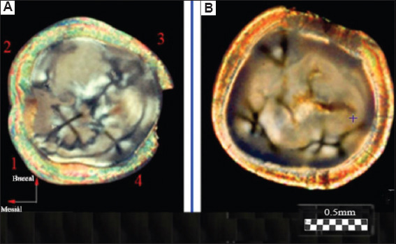
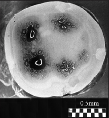
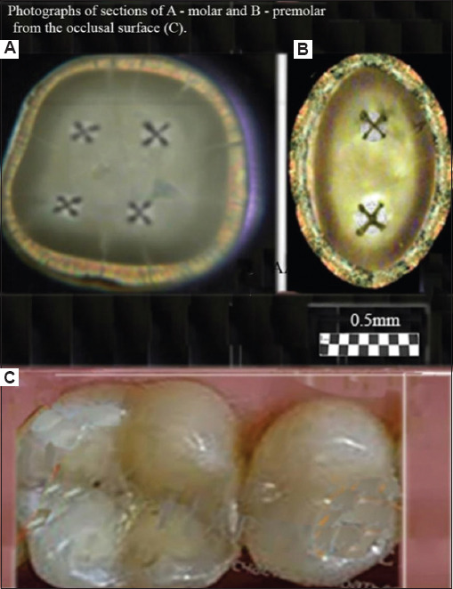
Results: The dentinal tubules were shown to originate from the center of the cusps in both molar and premolar teeth, forming S-shaped fiber bundles with axial-radial symmetry. These bundles were arranged along axes, extending from the pulp toward the centers of the cusps of the tooth crown. Within these zones, distinct optical patterns resembling conoscopic figures in the form of a "Maltese cross" were observed. This indicates a highly ordered architecture composed of optically anisotropic uniaxial tubules. The optical data were correlated well with findings obtained by electrometric and ion diffusion methods, including dentinal tubule staining.
Conclusion: The polarization optical is a valuable tool for studying various regional organizations of dentinal tubules in dentin.

 求助内容:
求助内容: 应助结果提醒方式:


