{"title":"A customized large single-piece bifrontal implant for post-craniectomy defect reconstruction: A case study.","authors":"Omid Ghaderzadeh, Ehsan Amirbeyk, Seyed Roholah Ghodsi, Zahra Namazi, Lobat Tayebi","doi":"10.14440/jbm.0019","DOIUrl":null,"url":null,"abstract":"<p><strong>Background: </strong>Large bifrontal defects pose unique reconstruction challenges due to their complex curvature and mechanical requirements. This case demonstrated how computer-aided design/manufacturing (CAD/CAM) enabled precise single-piece polymethyl methacrylate (PMMA) implant fabrication, thereby overcoming traditional limitations.</p><p><strong>Case presentation: </strong>A 25-year-old male who had undergone bifrontal decompressive craniectomy suffered a severe traumatic brain injury. The autologous bone flap had been temporarily stored in a subcutaneous fat area of the abdomen for 3 months to preserve its viability. A secondary cranioplasty was then performed using titanium miniplates and self-tapping screws for final fixation. After 2 years, the patient developed empyema and a brain abscess; the infected bone flap was removed. A skull computed tomography (CT) scan was conducted, and a prosthesis was created from PMMA by employing CAD. In the sagittal plane, the defect extended from the frontal bone and surpassed the coronal suture, while in the coronal plane, it reached the temporal region on both sides. The prosthesis was fabricated through rapid prototyping based on CT scan images. Surgery was performed using a patient-specific prosthesis that adequately covered the defect area. Facial aesthetics were restored, and no complications occurred. The patient was followed clinically and radiologically for 1 year, during which no postoperative complications or signs of implant-related issues were observed.</p><p><strong>Conclusion: </strong>This CAD/CAM single-piece PMMA implant successfully restored large bifrontal defects, suggesting that it may find broader applications in complex cranioplasties and could achieve improved outcomes.</p>","PeriodicalId":73618,"journal":{"name":"Journal of biological methods","volume":"12 3","pages":"e99010070"},"PeriodicalIF":0.0000,"publicationDate":"2025-08-12","publicationTypes":"Journal Article","fieldsOfStudy":null,"isOpenAccess":false,"openAccessPdf":"https://www.ncbi.nlm.nih.gov/pmc/articles/PMC12422115/pdf/","citationCount":"0","resultStr":null,"platform":"Semanticscholar","paperid":null,"PeriodicalName":"Journal of biological methods","FirstCategoryId":"1085","ListUrlMain":"https://doi.org/10.14440/jbm.0019","RegionNum":0,"RegionCategory":null,"ArticlePicture":[],"TitleCN":null,"AbstractTextCN":null,"PMCID":null,"EPubDate":"2025/1/1 0:00:00","PubModel":"eCollection","JCR":"","JCRName":"","Score":null,"Total":0}
引用次数: 0
Abstract
Background: Large bifrontal defects pose unique reconstruction challenges due to their complex curvature and mechanical requirements. This case demonstrated how computer-aided design/manufacturing (CAD/CAM) enabled precise single-piece polymethyl methacrylate (PMMA) implant fabrication, thereby overcoming traditional limitations.
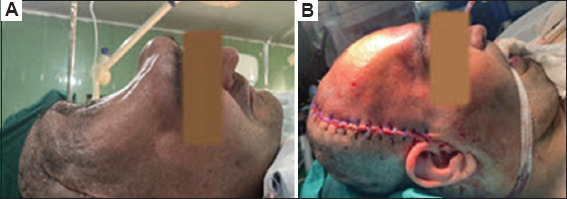
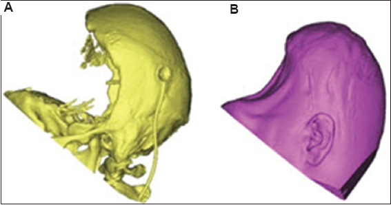
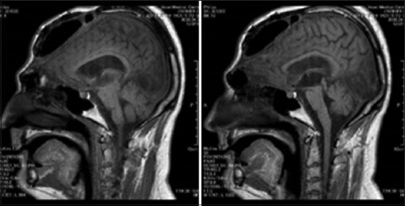
Case presentation: A 25-year-old male who had undergone bifrontal decompressive craniectomy suffered a severe traumatic brain injury. The autologous bone flap had been temporarily stored in a subcutaneous fat area of the abdomen for 3 months to preserve its viability. A secondary cranioplasty was then performed using titanium miniplates and self-tapping screws for final fixation. After 2 years, the patient developed empyema and a brain abscess; the infected bone flap was removed. A skull computed tomography (CT) scan was conducted, and a prosthesis was created from PMMA by employing CAD. In the sagittal plane, the defect extended from the frontal bone and surpassed the coronal suture, while in the coronal plane, it reached the temporal region on both sides. The prosthesis was fabricated through rapid prototyping based on CT scan images. Surgery was performed using a patient-specific prosthesis that adequately covered the defect area. Facial aesthetics were restored, and no complications occurred. The patient was followed clinically and radiologically for 1 year, during which no postoperative complications or signs of implant-related issues were observed.
Conclusion: This CAD/CAM single-piece PMMA implant successfully restored large bifrontal defects, suggesting that it may find broader applications in complex cranioplasties and could achieve improved outcomes.

 求助内容:
求助内容: 应助结果提醒方式:


