Moataz D Abouammo, Hossam S Elsherif, Medhat M H Mansour, Magdy E Saafan, Ricardo L Carrau, Mahmoud F Abdelaziz
{"title":"Assessment of endoscopic and external approaches for frontal sinus lesions.","authors":"Moataz D Abouammo, Hossam S Elsherif, Medhat M H Mansour, Magdy E Saafan, Ricardo L Carrau, Mahmoud F Abdelaziz","doi":"10.1002/wjo2.225","DOIUrl":null,"url":null,"abstract":"<p><strong>Objectives: </strong>The use of endoscopic approaches has revolutionized the management of frontal sinus (FS) lesions. However, external approaches still play a significant role in select conditions. Various factors determine the decision to utilize endoscopic or external approaches such as the lesion location, extension, and patient's characteristics. The study aims to define certain FS indices for accurate selection of the most suitable approach for each patient.</p><p><strong>Methods: </strong>A descriptive study was performed, based on endoscopic and external cadaveric dissections. Quantitative analyses including horizontal, anteroposterior diameters, and exposure area were performed for each approach using the navigation system. Patients with various FS lesions were included and their data were collected and evaluated.</p><p><strong>Results: </strong>Fifteen cadavers were analyzed. The average anteroposterior diameter on the midsagittal plane was 12.3 mm, distance from the midline to the lateralmost point was 21.8 mm on the right and 23.1 mm on the left side. The exposure area on the right side for Draf Ⅱa, and Draf Ⅱb were 64.6, 115.0 mm<sup>2</sup> while on the left side were 67.0, 125.0 mm<sup>2</sup>. For Draf Ⅲ, the exposure area was 377.0 mm<sup>2</sup>. A total of 41 patients were included in the clinical correlation.</p><p><strong>Conclusions: </strong>FS with a narrow anteroposterior diameter and longer horizontal diameter are difficult to access endoscopically, especially for lesions affecting the lateral recess of the sinus, and may require a combination with an external approach. FS approaches can be selected according to the sinus morphology of each patient, the surgeon's preferences, institutional resources, and the lesion's nature and extension.</p>","PeriodicalId":32097,"journal":{"name":"World Journal of OtorhinolaryngologyHead and Neck Surgery","volume":"11 3","pages":"375-384"},"PeriodicalIF":1.4000,"publicationDate":"2024-12-11","publicationTypes":"Journal Article","fieldsOfStudy":null,"isOpenAccess":false,"openAccessPdf":"https://www.ncbi.nlm.nih.gov/pmc/articles/PMC12418322/pdf/","citationCount":"0","resultStr":null,"platform":"Semanticscholar","paperid":null,"PeriodicalName":"World Journal of OtorhinolaryngologyHead and Neck Surgery","FirstCategoryId":"3","ListUrlMain":"https://doi.org/10.1002/wjo2.225","RegionNum":0,"RegionCategory":null,"ArticlePicture":[],"TitleCN":null,"AbstractTextCN":null,"PMCID":null,"EPubDate":"2025/9/1 0:00:00","PubModel":"eCollection","JCR":"Q2","JCRName":"Medicine","Score":null,"Total":0}
引用次数: 0
Abstract
Objectives: The use of endoscopic approaches has revolutionized the management of frontal sinus (FS) lesions. However, external approaches still play a significant role in select conditions. Various factors determine the decision to utilize endoscopic or external approaches such as the lesion location, extension, and patient's characteristics. The study aims to define certain FS indices for accurate selection of the most suitable approach for each patient.
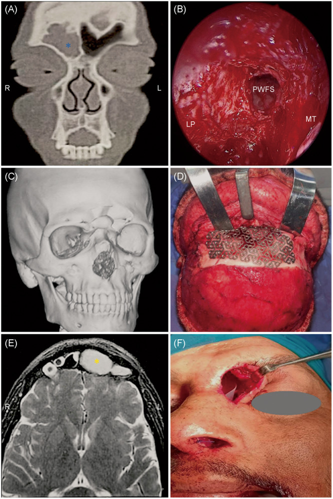
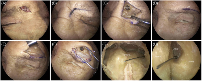
Methods: A descriptive study was performed, based on endoscopic and external cadaveric dissections. Quantitative analyses including horizontal, anteroposterior diameters, and exposure area were performed for each approach using the navigation system. Patients with various FS lesions were included and their data were collected and evaluated.
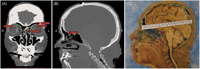
Results: Fifteen cadavers were analyzed. The average anteroposterior diameter on the midsagittal plane was 12.3 mm, distance from the midline to the lateralmost point was 21.8 mm on the right and 23.1 mm on the left side. The exposure area on the right side for Draf Ⅱa, and Draf Ⅱb were 64.6, 115.0 mm2 while on the left side were 67.0, 125.0 mm2. For Draf Ⅲ, the exposure area was 377.0 mm2. A total of 41 patients were included in the clinical correlation.
Conclusions: FS with a narrow anteroposterior diameter and longer horizontal diameter are difficult to access endoscopically, especially for lesions affecting the lateral recess of the sinus, and may require a combination with an external approach. FS approaches can be selected according to the sinus morphology of each patient, the surgeon's preferences, institutional resources, and the lesion's nature and extension.
 求助内容:
求助内容: 应助结果提醒方式:


