{"title":"Rare case of extrahepatic biliary obstruction caused by a duodenal foreign body in a cat.","authors":"Hyunwook Myung, Ilyong Yun, Jonghyop Lee","doi":"10.1177/20551169251366439","DOIUrl":null,"url":null,"abstract":"<p><strong>Case summary: </strong>A spayed female British Shorthair cat aged 1 year and weighing 2.6 kg presented with a 5-day history of vomiting and anorexia. Physical examination revealed icterus, and serum biochemistry showed markedly elevated bilirubin and liver enzyme levels. Abdominal ultrasonography revealed a dilated common bile duct (4 mm), mild gallbladder wall thickening and a round, hyperechoic foreign body located at the major duodenal papilla. A contrast-enhanced CT scan confirmed the presence of a 1.9 cm doughnut-shaped foreign body in the proximal descending duodenum, causing extramural compression of the common bile duct. Exploratory laparotomy and enterotomy were performed to remove the object. The cat recovered uneventfully, with normalisation of biliary parameters within 48 h and resolution of clinical signs by postoperative day 4.</p><p><strong>Relevance and novel information: </strong>This case highlights a rare but surgically treatable cause of extrahepatic biliary obstruction (EHBO) in cats caused by a duodenal foreign body exerting extraluminal compression without intraluminal migration or mucosal invasion. It emphasises the value of cross-sectional imaging and timely surgical intervention in achieving favourable outcomes. To the authors' knowledge, this is the first peer-reviewed report of feline EHBO caused by extramural duodenal compression that was successfully resolved without biliary tract incision.</p>","PeriodicalId":36588,"journal":{"name":"Journal of Feline Medicine and Surgery Open Reports","volume":"11 2","pages":"20551169251366439"},"PeriodicalIF":0.7000,"publicationDate":"2025-09-07","publicationTypes":"Journal Article","fieldsOfStudy":null,"isOpenAccess":false,"openAccessPdf":"https://www.ncbi.nlm.nih.gov/pmc/articles/PMC12417643/pdf/","citationCount":"0","resultStr":null,"platform":"Semanticscholar","paperid":null,"PeriodicalName":"Journal of Feline Medicine and Surgery Open Reports","FirstCategoryId":"1085","ListUrlMain":"https://doi.org/10.1177/20551169251366439","RegionNum":0,"RegionCategory":null,"ArticlePicture":[],"TitleCN":null,"AbstractTextCN":null,"PMCID":null,"EPubDate":"2025/7/1 0:00:00","PubModel":"eCollection","JCR":"Q3","JCRName":"VETERINARY SCIENCES","Score":null,"Total":0}
引用次数: 0
Abstract
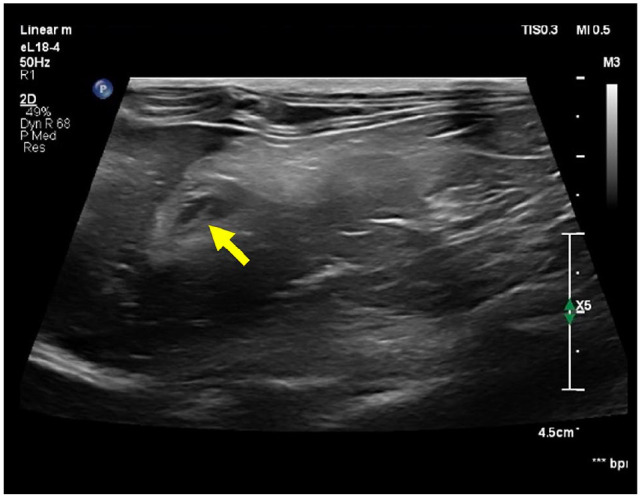
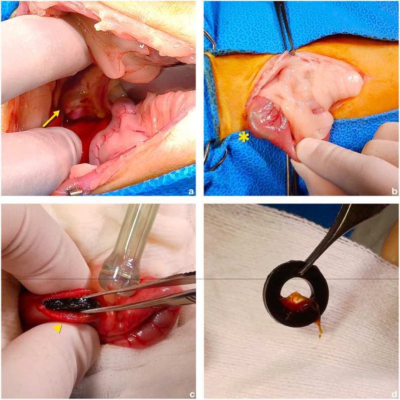
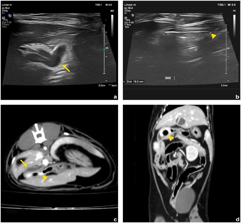
Case summary: A spayed female British Shorthair cat aged 1 year and weighing 2.6 kg presented with a 5-day history of vomiting and anorexia. Physical examination revealed icterus, and serum biochemistry showed markedly elevated bilirubin and liver enzyme levels. Abdominal ultrasonography revealed a dilated common bile duct (4 mm), mild gallbladder wall thickening and a round, hyperechoic foreign body located at the major duodenal papilla. A contrast-enhanced CT scan confirmed the presence of a 1.9 cm doughnut-shaped foreign body in the proximal descending duodenum, causing extramural compression of the common bile duct. Exploratory laparotomy and enterotomy were performed to remove the object. The cat recovered uneventfully, with normalisation of biliary parameters within 48 h and resolution of clinical signs by postoperative day 4.
Relevance and novel information: This case highlights a rare but surgically treatable cause of extrahepatic biliary obstruction (EHBO) in cats caused by a duodenal foreign body exerting extraluminal compression without intraluminal migration or mucosal invasion. It emphasises the value of cross-sectional imaging and timely surgical intervention in achieving favourable outcomes. To the authors' knowledge, this is the first peer-reviewed report of feline EHBO caused by extramural duodenal compression that was successfully resolved without biliary tract incision.

 求助内容:
求助内容: 应助结果提醒方式:


