Amandeep Singh, Manmohan Singh, Salavat R Aglyamov, David Mayerich, Kirill V Larin
{"title":"Quantifying age and spatial variations of bone marrow elasticity with noncontact optical coherence elastography.","authors":"Amandeep Singh, Manmohan Singh, Salavat R Aglyamov, David Mayerich, Kirill V Larin","doi":"10.1117/1.JBO.30.12.124505","DOIUrl":null,"url":null,"abstract":"<p><strong>Significance: </strong>The bone marrow is essential in immune regulation to maintain body homeostasis and to control the trafficking of stromal cells. A framework of connective tissue upholds bone marrow cells to maintain their mechanical and functional integrity. The biomechanical characterization of the bone marrow may provide useful insights for diagnosing hematologic diseases such as primary myelofibrosis. Optical coherence elastography (OCE) can measure the mechanical properties of tissues with high spatiotemporal resolution and may be well-suited for characterizing bone marrow elasticity.</p><p><strong>Aim: </strong>We demonstrate the quantification of the elastic modulus of bone marrow <i>ex vivo</i> at different locations along the diaphysis of mice femurs and compare the elastic modulus within different age groups of mice femurs.</p><p><strong>Approach: </strong>The femur bone marrow of CD1 mice, <math><mrow><mo>∼</mo> <mn>12</mn></mrow> </math> weeks old (young adult), 24 weeks old (mature adult), and 1 year old (old adult), was imaged with OCE ( <math><mrow><mi>N</mi> <mo>=</mo> <mn>4</mn></mrow> </math> femurs for each age group) to investigate the change in stiffness with age and location along the femur. A noncontact air-coupled ultrasound (ACUS) transducer induced elastic waves in the bone marrow, which were detected by phase-sensitive optical coherence tomography. The ACUS-OCE measurements were taken at three different locations along the diaphysis from the proximal end to the distal end to investigate the spatial stiffness variations.</p><p><strong>Results: </strong>The results show that the stiffness of femoral bone marrow increases significantly with age ( <math><mrow><mi>p</mi> <mo><</mo> <mn>0.001</mn></mrow> </math> ), but there was no significant difference in Young's moduli among the locations for young ( <math> <mrow> <msup><mrow><mi>χ</mi></mrow> <mrow><mn>2</mn></mrow> </msup> <mo>(</mo> <mn>2</mn> <mo>)</mo> <mo>=</mo> <mn>2.15</mn></mrow> </math> , <math><mrow><mi>p</mi> <mo>=</mo> <mn>0.33</mn></mrow> </math> ), mature ( <math> <mrow> <msup><mrow><mi>χ</mi></mrow> <mrow><mn>2</mn></mrow> </msup> <mo>(</mo> <mn>2</mn> <mo>)</mo> <mo>=</mo> <mn>5.68</mn></mrow> </math> , <math><mrow><mi>p</mi> <mo>=</mo> <mn>0.058</mn></mrow> </math> ), and old ( <math> <mrow> <msup><mrow><mi>χ</mi></mrow> <mrow><mn>2</mn></mrow> </msup> <mo>(</mo> <mn>2</mn> <mo>)</mo> <mo>=</mo> <mn>5.73</mn></mrow> </math> , <math><mrow><mi>p</mi> <mo>=</mo> <mn>0.056</mn></mrow> </math> ) mice femur samples.</p><p><strong>Conclusions: </strong>These findings show that OCE is promising for mapping the stiffness of the intact bone marrow and could be used for minimally invasive clinical applications.</p>","PeriodicalId":15264,"journal":{"name":"Journal of Biomedical Optics","volume":"30 12","pages":"124505"},"PeriodicalIF":2.9000,"publicationDate":"2025-12-01","publicationTypes":"Journal Article","fieldsOfStudy":null,"isOpenAccess":false,"openAccessPdf":"https://www.ncbi.nlm.nih.gov/pmc/articles/PMC12422286/pdf/","citationCount":"0","resultStr":null,"platform":"Semanticscholar","paperid":null,"PeriodicalName":"Journal of Biomedical Optics","FirstCategoryId":"3","ListUrlMain":"https://doi.org/10.1117/1.JBO.30.12.124505","RegionNum":3,"RegionCategory":"医学","ArticlePicture":[],"TitleCN":null,"AbstractTextCN":null,"PMCID":null,"EPubDate":"2025/9/10 0:00:00","PubModel":"Epub","JCR":"Q2","JCRName":"BIOCHEMICAL RESEARCH METHODS","Score":null,"Total":0}
引用次数: 0
Abstract
Significance: The bone marrow is essential in immune regulation to maintain body homeostasis and to control the trafficking of stromal cells. A framework of connective tissue upholds bone marrow cells to maintain their mechanical and functional integrity. The biomechanical characterization of the bone marrow may provide useful insights for diagnosing hematologic diseases such as primary myelofibrosis. Optical coherence elastography (OCE) can measure the mechanical properties of tissues with high spatiotemporal resolution and may be well-suited for characterizing bone marrow elasticity.
Aim: We demonstrate the quantification of the elastic modulus of bone marrow ex vivo at different locations along the diaphysis of mice femurs and compare the elastic modulus within different age groups of mice femurs.
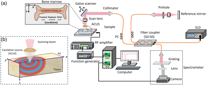
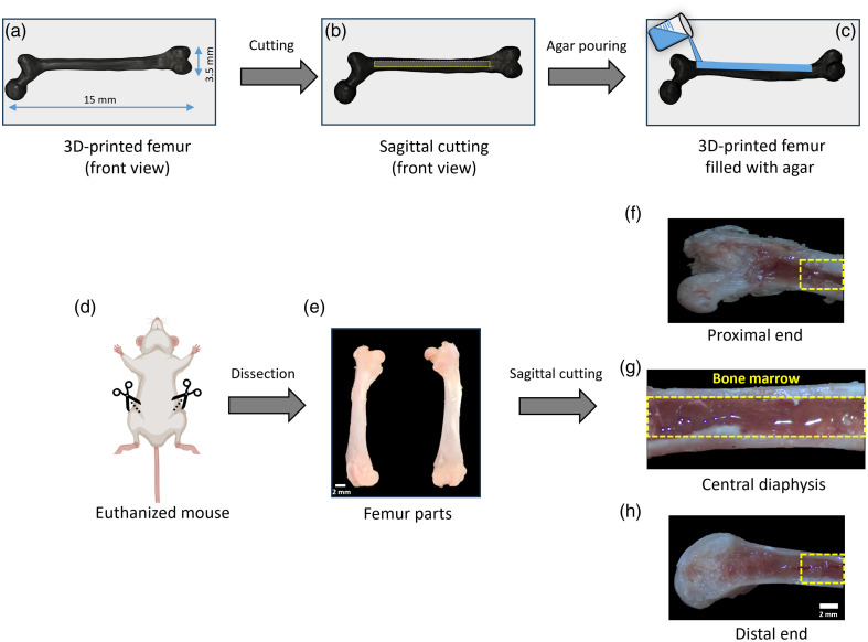
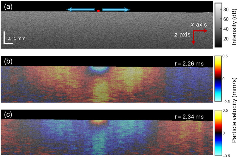
Approach: The femur bone marrow of CD1 mice, weeks old (young adult), 24 weeks old (mature adult), and 1 year old (old adult), was imaged with OCE ( femurs for each age group) to investigate the change in stiffness with age and location along the femur. A noncontact air-coupled ultrasound (ACUS) transducer induced elastic waves in the bone marrow, which were detected by phase-sensitive optical coherence tomography. The ACUS-OCE measurements were taken at three different locations along the diaphysis from the proximal end to the distal end to investigate the spatial stiffness variations.
Results: The results show that the stiffness of femoral bone marrow increases significantly with age ( ), but there was no significant difference in Young's moduli among the locations for young ( , ), mature ( , ), and old ( , ) mice femur samples.
Conclusions: These findings show that OCE is promising for mapping the stiffness of the intact bone marrow and could be used for minimally invasive clinical applications.
期刊介绍:
The Journal of Biomedical Optics publishes peer-reviewed papers on the use of modern optical technology for improved health care and biomedical research.

 求助内容:
求助内容: 应助结果提醒方式:


