Marcelo Klotz Dall'Agnol, Mateus Bond Boghossian, André Orsini Ardengh, Ygor Rocha Fernandes, Matheus de Oliveira Veras, Evellin Souza Valentim Dos Santos, Tomazo Antonio Prince Franzini, Wanderley Marques Bernardo, Eduardo Guimarães Hourneaux de Moura
{"title":"Peroral cholangioscopy for detecting residual stones missed by cholangiography: Systematic review and meta-analysis.","authors":"Marcelo Klotz Dall'Agnol, Mateus Bond Boghossian, André Orsini Ardengh, Ygor Rocha Fernandes, Matheus de Oliveira Veras, Evellin Souza Valentim Dos Santos, Tomazo Antonio Prince Franzini, Wanderley Marques Bernardo, Eduardo Guimarães Hourneaux de Moura","doi":"10.1055/a-2676-4062","DOIUrl":null,"url":null,"abstract":"<p><strong>Background and study aims: </strong>Residual bile duct stones may persist despite negative cholangiographic findings after endoscopic retrograde cholangiopancreatography, increasing risk of recurrence and complications. This systematic review and meta-analysis aimed to determine the detection rate of residual stones identified by peroral cholangioscopy (POC), alongside stone characteristics and baseline patient features.</p><p><strong>Methods: </strong>A comprehensive search was conducted in MEDLINE, Cochrane Library, EMBASE, and LILACS through August 2024. Eligible studies included patients undergoing POC after negative occlusion cholangiography. The primary outcome was the pooled residual stone detection rate. Secondary outcomes included residual stone characteristics, adverse events (AEs), and baseline clinical parameters. Subgroup analysis was performed according to cholangioscopy technique used.</p><p><strong>Results: </strong>Nine studies comprising 485 procedures were included. The pooled residual stone detection rate was 27% (95% confidence interval 23%-31%), with higher detection using digital single-operator cholangioscopy (32%) compared with direct peroral cholangioscopy (25%) and Mother-Baby systems (24%). Residual stones had a mean size of 4.51 mm, with an average of 1.55 stones per positive procedure. Mild AEs occurred in 3% of cases, with no serious complications reported. Baseline characteristics showed an average initial stone size of 12.89 mm, a mean common bile duct diameter of 15.28 mm, and lithotripsy use in 57% of cases.</p><p><strong>Conclusions: </strong>POC identified residual stones in over one-fourth of patients following negative cholangiography. Detection rates were highest with digital systems. The procedure demonstrated a strong safety profile and may play an important role in confirming complete ductal clearance.</p>","PeriodicalId":11671,"journal":{"name":"Endoscopy International Open","volume":"13 ","pages":"a26764062"},"PeriodicalIF":2.3000,"publicationDate":"2025-08-26","publicationTypes":"Journal Article","fieldsOfStudy":null,"isOpenAccess":false,"openAccessPdf":"https://www.ncbi.nlm.nih.gov/pmc/articles/PMC12417788/pdf/","citationCount":"0","resultStr":null,"platform":"Semanticscholar","paperid":null,"PeriodicalName":"Endoscopy International Open","FirstCategoryId":"1085","ListUrlMain":"https://doi.org/10.1055/a-2676-4062","RegionNum":0,"RegionCategory":null,"ArticlePicture":[],"TitleCN":null,"AbstractTextCN":null,"PMCID":null,"EPubDate":"2025/1/1 0:00:00","PubModel":"eCollection","JCR":"Q3","JCRName":"GASTROENTEROLOGY & HEPATOLOGY","Score":null,"Total":0}
引用次数: 0
Abstract
Background and study aims: Residual bile duct stones may persist despite negative cholangiographic findings after endoscopic retrograde cholangiopancreatography, increasing risk of recurrence and complications. This systematic review and meta-analysis aimed to determine the detection rate of residual stones identified by peroral cholangioscopy (POC), alongside stone characteristics and baseline patient features.
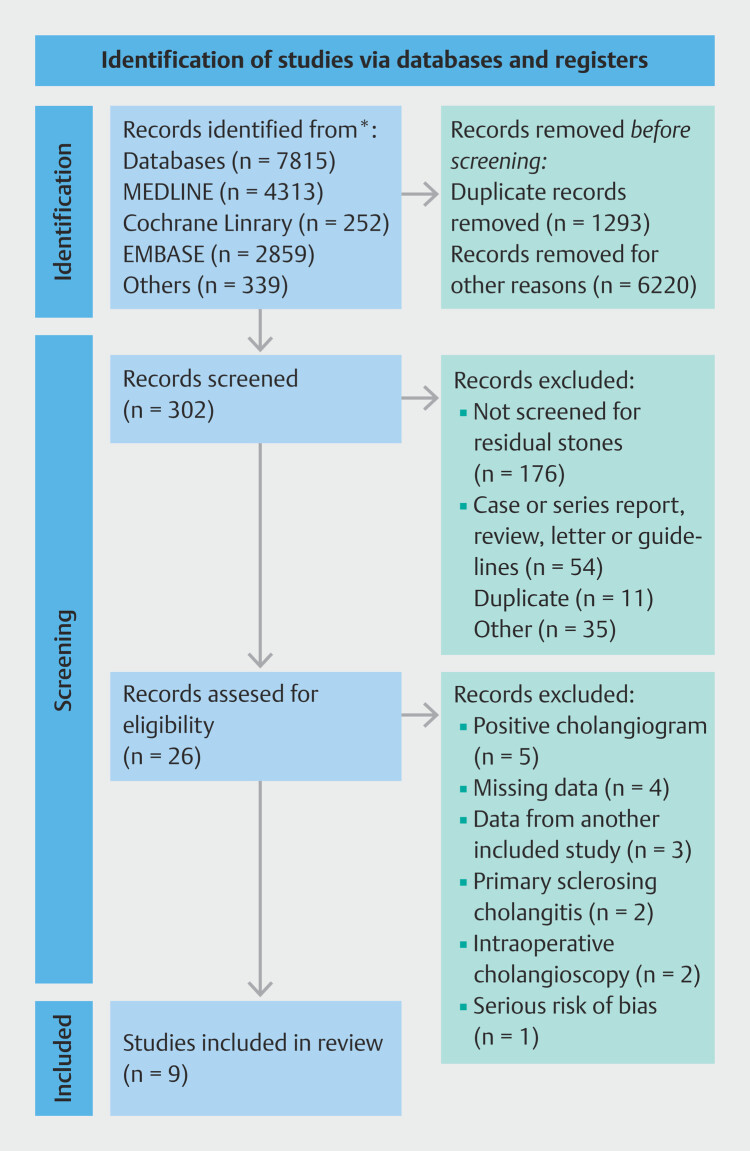
Methods: A comprehensive search was conducted in MEDLINE, Cochrane Library, EMBASE, and LILACS through August 2024. Eligible studies included patients undergoing POC after negative occlusion cholangiography. The primary outcome was the pooled residual stone detection rate. Secondary outcomes included residual stone characteristics, adverse events (AEs), and baseline clinical parameters. Subgroup analysis was performed according to cholangioscopy technique used.
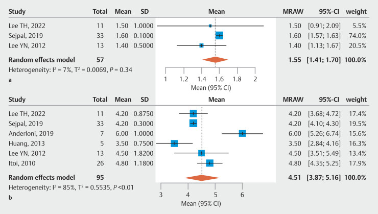
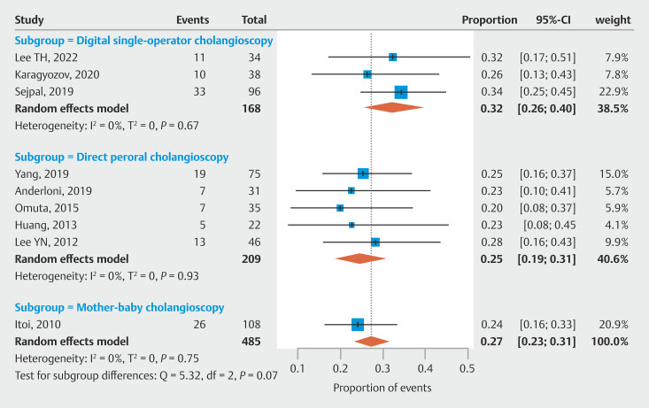
Results: Nine studies comprising 485 procedures were included. The pooled residual stone detection rate was 27% (95% confidence interval 23%-31%), with higher detection using digital single-operator cholangioscopy (32%) compared with direct peroral cholangioscopy (25%) and Mother-Baby systems (24%). Residual stones had a mean size of 4.51 mm, with an average of 1.55 stones per positive procedure. Mild AEs occurred in 3% of cases, with no serious complications reported. Baseline characteristics showed an average initial stone size of 12.89 mm, a mean common bile duct diameter of 15.28 mm, and lithotripsy use in 57% of cases.
Conclusions: POC identified residual stones in over one-fourth of patients following negative cholangiography. Detection rates were highest with digital systems. The procedure demonstrated a strong safety profile and may play an important role in confirming complete ductal clearance.

 求助内容:
求助内容: 应助结果提醒方式:


