Differential cortical hemodynamics during standard and reversed visually guided navigation: An fNIRS-based investigation
IF 3.3
3区 医学
Q2 CLINICAL NEUROLOGY
引用次数: 0
Abstract
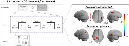
Visuospatial perception, which is based on the comprehension of objects and space, requires spatial attention to the surrounding environment. Stimulus-related elements that affect visuospatial tasks include object geometry, familiarity, complexity, and picture plane versus depth rotation. The dorsal stream pathway from the visual cortex, which is implicated in spatial processing, reflects the spatial component needed to orient the focus of attention to the location of the expected target stimulus. It is activated during spatial localization. While processing spatial information, visual, somatosensory, and auditory information is received from the inferotemporal cortex, medial and superior parietal cortices, and transverse temporal gyrus, and is projected directly toward the prefrontal cortex, which includes the premotor cortex. In this study 10 volunteers performed standard and reverse visually guided weight-shifting training tasks. This study aimed to investigate the hemodynamic response of the parietal to occipital cortex during these tasks using a 41-channel functional near-infrared spectroscopy system. During the standard navigation task, the right supramarginal gyrus showed a significant increase in oxy-hemoglobin (HbO) and total-hemoglobin (HbT) values. In contrast, the reverse navigation task showed significant increments in HbO values in the right angular gyrus (AG) and left somatosensory association cortex (SAC) and in HbT values in the left SAC and both AG. Thus, according to our results, spatial processing based on reversal may be different. Moreover, a difference in the amount of oxygen was observed. Further studies are required to understand the activated neural mechanisms when sensory inputs differ during spatial information processing.
在标准和反向视觉引导导航中的皮质血流动力学差异:一项基于fnir的研究。
视觉空间感知以对物体和空间的理解为基础,需要对周围环境进行空间关注。影响视觉空间任务的刺激相关元素包括物体几何、熟悉度、复杂性和画面平面与深度旋转。来自视觉皮层的背侧流通路与空间加工有关,反映了将注意力焦点定向到预期目标刺激位置所需的空间成分。它在空间定位过程中被激活。在处理空间信息时,视觉、体感和听觉信息从颞下皮层、顶叶内侧和顶叶上部皮层以及颞横回接收,并直接投射到包括前运动皮层在内的前额叶皮层。在这项研究中,10名志愿者完成了标准和反向视觉引导的体重转移训练任务。本研究旨在利用41通道功能近红外光谱系统研究这些任务中顶叶到枕叶皮层的血流动力学反应。在标准的导航任务中,右侧边缘上回的氧血红蛋白(HbO)和总血红蛋白(HbT)值显著增加。相反,反向导航任务显示右侧角回(AG)和左侧体感关联皮层(SAC)的HbO值显著增加,左侧SAC和两个AG的HbT值显著增加。因此,根据我们的结果,基于反转的空间处理可能会有所不同。此外,还观察到氧气量的差异。在空间信息处理过程中,当感觉输入不同时,被激活的神经机制需要进一步的研究。
本文章由计算机程序翻译,如有差异,请以英文原文为准。
求助全文
约1分钟内获得全文
求助全文
来源期刊
Journal of Neuroradiology
医学-核医学
CiteScore
6.10
自引率
5.70%
发文量
142
审稿时长
6-12 weeks
期刊介绍:
The Journal of Neuroradiology is a peer-reviewed journal, publishing worldwide clinical and basic research in the field of diagnostic and Interventional neuroradiology, translational and molecular neuroimaging, and artificial intelligence in neuroradiology.
The Journal of Neuroradiology considers for publication articles, reviews, technical notes and letters to the editors (correspondence section), provided that the methodology and scientific content are of high quality, and that the results will have substantial clinical impact and/or physiological importance.
 求助内容:
求助内容: 应助结果提醒方式:


