John M Fender, Amy M Habing, Layla K Behbahani, Sean H McCready
{"title":"A New Wave of Ultrasound Phantoms Using Real Fixed Organs: Birth of the Danny Phantom.","authors":"John M Fender, Amy M Habing, Layla K Behbahani, Sean H McCready","doi":"10.1111/vru.70078","DOIUrl":null,"url":null,"abstract":"<p><p>Ultrasonography in veterinary medicine serves a vital role in the diagnosis and management of various medical conditions by allowing noninvasive visualization of internal structures. Veterinary students face many challenges in gaining hands-on experience with ultrasound equipment and developing competencies in ultrasonography. This is largely due to the limited access and ethical dilemmas of live animal models and the high cost of commercial phantoms. To solve these issues, the niche of cost-effective amateur models has exponentially increased. However, while these at-home models solve the financial issues associated with commercial phantoms, they still lack the realism and fidelity necessary to simulate the real-time feedback needed to gain the spatial awareness of this dynamic imaging modality. To foster successful day-one-ready veterinary students, The Ohio State University College of Veterinary Medicine acknowledged that a better solution should be possible. A prospective anatomic study was performed to recognize the imaging anatomy and usability of a new model termed the Danny Phantom. This model was developed by testing various amateur phantom materials from both the literature and self-discovered. These materials were analyzed and deemed satisfactory versus unsatisfactory based on fulfillment of predetermined criteria of an ideal phantom model. It was determined that real fixed organs can be encased in traditional bovine gelatin to produce an ultrasound phantom with recognizable parenchyma. Other additives can be included to give the phantom an imitated peritoneal space and prevent spoilage of the gelatin for an extended period of time.</p>","PeriodicalId":23581,"journal":{"name":"Veterinary Radiology & Ultrasound","volume":"66 5","pages":"e70078"},"PeriodicalIF":1.5000,"publicationDate":"2025-09-01","publicationTypes":"Journal Article","fieldsOfStudy":null,"isOpenAccess":false,"openAccessPdf":"https://www.ncbi.nlm.nih.gov/pmc/articles/PMC12423588/pdf/","citationCount":"0","resultStr":null,"platform":"Semanticscholar","paperid":null,"PeriodicalName":"Veterinary Radiology & Ultrasound","FirstCategoryId":"97","ListUrlMain":"https://doi.org/10.1111/vru.70078","RegionNum":2,"RegionCategory":"农林科学","ArticlePicture":[],"TitleCN":null,"AbstractTextCN":null,"PMCID":null,"EPubDate":"","PubModel":"","JCR":"Q2","JCRName":"VETERINARY SCIENCES","Score":null,"Total":0}
引用次数: 0
Abstract
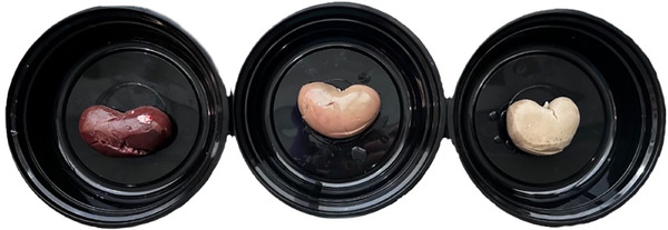
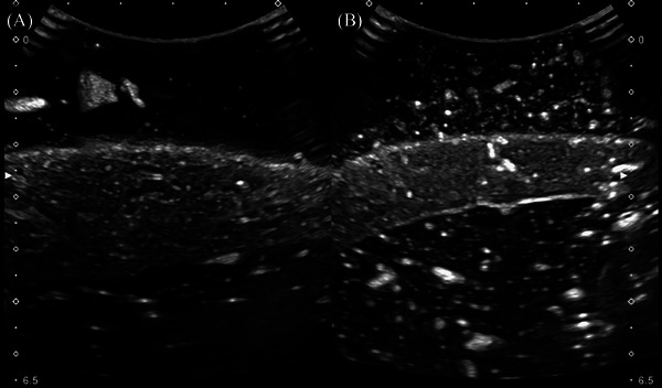
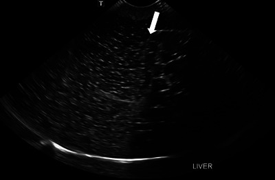
Ultrasonography in veterinary medicine serves a vital role in the diagnosis and management of various medical conditions by allowing noninvasive visualization of internal structures. Veterinary students face many challenges in gaining hands-on experience with ultrasound equipment and developing competencies in ultrasonography. This is largely due to the limited access and ethical dilemmas of live animal models and the high cost of commercial phantoms. To solve these issues, the niche of cost-effective amateur models has exponentially increased. However, while these at-home models solve the financial issues associated with commercial phantoms, they still lack the realism and fidelity necessary to simulate the real-time feedback needed to gain the spatial awareness of this dynamic imaging modality. To foster successful day-one-ready veterinary students, The Ohio State University College of Veterinary Medicine acknowledged that a better solution should be possible. A prospective anatomic study was performed to recognize the imaging anatomy and usability of a new model termed the Danny Phantom. This model was developed by testing various amateur phantom materials from both the literature and self-discovered. These materials were analyzed and deemed satisfactory versus unsatisfactory based on fulfillment of predetermined criteria of an ideal phantom model. It was determined that real fixed organs can be encased in traditional bovine gelatin to produce an ultrasound phantom with recognizable parenchyma. Other additives can be included to give the phantom an imitated peritoneal space and prevent spoilage of the gelatin for an extended period of time.
期刊介绍:
Veterinary Radiology & Ultrasound is a bimonthly, international, peer-reviewed, research journal devoted to the fields of veterinary diagnostic imaging and radiation oncology. Established in 1958, it is owned by the American College of Veterinary Radiology and is also the official journal for six affiliate veterinary organizations. Veterinary Radiology & Ultrasound is represented on the International Committee of Medical Journal Editors, World Association of Medical Editors, and Committee on Publication Ethics.
The mission of Veterinary Radiology & Ultrasound is to serve as a leading resource for high quality articles that advance scientific knowledge and standards of clinical practice in the areas of veterinary diagnostic radiology, computed tomography, magnetic resonance imaging, ultrasonography, nuclear imaging, radiation oncology, and interventional radiology. Manuscript types include original investigations, imaging diagnosis reports, review articles, editorials and letters to the Editor. Acceptance criteria include originality, significance, quality, reader interest, composition and adherence to author guidelines.

 求助内容:
求助内容: 应助结果提醒方式:


