Giuliana de Campos Chaves Lamarque, Roberta Duarte Leme, Luciano Aparecido de Almeida Junior, Marília Pacifico Lucisano, Karina Fittipaldi Bombonato-Prado, Raquel Assed Bezerra Segato, Anne George, Francisco Wanderley Garcia Paula-Silva
{"title":"Effects triggered by tumor necrosis factor-α in immortalized murine dental pulp and pre-osteoblastic cells.","authors":"Giuliana de Campos Chaves Lamarque, Roberta Duarte Leme, Luciano Aparecido de Almeida Junior, Marília Pacifico Lucisano, Karina Fittipaldi Bombonato-Prado, Raquel Assed Bezerra Segato, Anne George, Francisco Wanderley Garcia Paula-Silva","doi":"10.1590/1807-3107bor-2025.vol39.087","DOIUrl":null,"url":null,"abstract":"<p><p>Tumor necrosis factor-alpha (TNF-α) is a cytokine involved in the immune-inflammatory response. It can induce an odontoblastic phenotype and enhance biomineralization in dental pulp mesenchymal stem cells but does not have the same effect on osteoblasts. The reasons for this differential response, despite the shared lineage of these cell types, are not yet clear. This study examined the effects of TNF-α on immortalized mouse dental pulp stem cells (OD-21) and pre-osteoblastic cells (MC3T3-E1). Cells were treated with recombinant TNF-α at concentrations of 1, 10, and 100 ng/mL. Cell viability, proliferation, and migration were assessed using the MTT, CyQUANT, and wound healing assays, respectively. Gene expression was assessed via real-time RT-PCR, and biomineralization was evaluated using alizarin red staining. Statistical analysis was conducted using one-way ANOVA followed by Tukey's post-hoc test (α = 0.05). TNF-α did not affect cell viability at any concentration (p > 0.05). Proliferation and migration increased after 12 h, with near-complete wound closure by 24 h. TNF-α promoted proliferation and migration in both cell types. OD-21 cells exhibited high levels of Tnfr1 and Runx2 expression and showed biomineralization. In contrast, MC3T3-E1 cells showed high Tnfr2 levels, suppressed Runx2, and inhibited biomineralization. These results highlight how TNF-α influences different cell types from the same lineage in distinct ways.</p>","PeriodicalId":9240,"journal":{"name":"Brazilian oral research","volume":"39 ","pages":"e087"},"PeriodicalIF":1.3000,"publicationDate":"2025-09-08","publicationTypes":"Journal Article","fieldsOfStudy":null,"isOpenAccess":false,"openAccessPdf":"https://www.ncbi.nlm.nih.gov/pmc/articles/PMC12419187/pdf/","citationCount":"0","resultStr":null,"platform":"Semanticscholar","paperid":null,"PeriodicalName":"Brazilian oral research","FirstCategoryId":"3","ListUrlMain":"https://doi.org/10.1590/1807-3107bor-2025.vol39.087","RegionNum":4,"RegionCategory":"医学","ArticlePicture":[],"TitleCN":null,"AbstractTextCN":null,"PMCID":null,"EPubDate":"2025/1/1 0:00:00","PubModel":"eCollection","JCR":"Q3","JCRName":"DENTISTRY, ORAL SURGERY & MEDICINE","Score":null,"Total":0}
引用次数: 0
Abstract
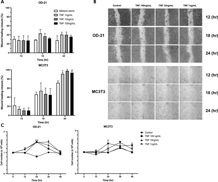
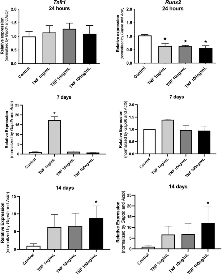
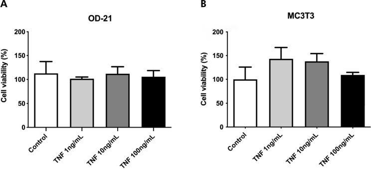
Tumor necrosis factor-alpha (TNF-α) is a cytokine involved in the immune-inflammatory response. It can induce an odontoblastic phenotype and enhance biomineralization in dental pulp mesenchymal stem cells but does not have the same effect on osteoblasts. The reasons for this differential response, despite the shared lineage of these cell types, are not yet clear. This study examined the effects of TNF-α on immortalized mouse dental pulp stem cells (OD-21) and pre-osteoblastic cells (MC3T3-E1). Cells were treated with recombinant TNF-α at concentrations of 1, 10, and 100 ng/mL. Cell viability, proliferation, and migration were assessed using the MTT, CyQUANT, and wound healing assays, respectively. Gene expression was assessed via real-time RT-PCR, and biomineralization was evaluated using alizarin red staining. Statistical analysis was conducted using one-way ANOVA followed by Tukey's post-hoc test (α = 0.05). TNF-α did not affect cell viability at any concentration (p > 0.05). Proliferation and migration increased after 12 h, with near-complete wound closure by 24 h. TNF-α promoted proliferation and migration in both cell types. OD-21 cells exhibited high levels of Tnfr1 and Runx2 expression and showed biomineralization. In contrast, MC3T3-E1 cells showed high Tnfr2 levels, suppressed Runx2, and inhibited biomineralization. These results highlight how TNF-α influences different cell types from the same lineage in distinct ways.
 求助内容:
求助内容: 应助结果提醒方式:


