Maha Ibrahim Metwally, Yassir Edrees Almalki, Marwa Fathy Khalil, Ahmed Mohamed Alsowey, Hazem Ibrahim Aly Tantawy, Mohamed Gaber Hamed, Shimaa Abdelmoneem, Sharifa Khalid Alduraibi, Ziyad A Almushayti, Shaker Hassan S Alshehri, Ahmed M Abdelkhalik Basha, Mohammad Abd Alkhalik Basha
{"title":"Quantitative MRI Dixon signal drop and fat fraction for differentiating bone marrow lesions: a two-center prospective analysis.","authors":"Maha Ibrahim Metwally, Yassir Edrees Almalki, Marwa Fathy Khalil, Ahmed Mohamed Alsowey, Hazem Ibrahim Aly Tantawy, Mohamed Gaber Hamed, Shimaa Abdelmoneem, Sharifa Khalid Alduraibi, Ziyad A Almushayti, Shaker Hassan S Alshehri, Ahmed M Abdelkhalik Basha, Mohammad Abd Alkhalik Basha","doi":"10.1186/s41747-025-00615-9","DOIUrl":null,"url":null,"abstract":"<p><strong>Background: </strong>Bone marrow (BM) lesion differentiation remains challenging, and quantitative magnetic resonance imaging (MRI) may enhance accuracy over conventional methods. We evaluated the diagnostic value and inter-reader reliability of Dixon-based signal drop (%drop) and fat fraction percentage (%fat) as adjuncts to existing protocols.</p><p><strong>Materials and methods: </strong>In this prospective two-center study, 172 patients with BM signal abnormalities underwent standardized 1.5-T MRI protocols, including Dixon sequences. Two musculoskeletal radiologists independently evaluated images and performed quantitative measurements of %drop and %fat. Final diagnoses were established through histopathology (n = 96) or imaging follow-up (n = 76). Diagnostic value was assessed using area under the receiver operating characteristic curve (AUROC), inter-reader reliability using Cohen's κ coefficient.</p><p><strong>Results: </strong>The consensus optimal cutoff was for %drop ≤ 19.8%, yielding 87.2% accuracy, 95.3% sensitivity, and 73.8% specificity, and that for %fat was ≤ 18.3%, achieving 86.6% accuracy, 96.3% sensitivity, and 70.8% specificity. Both metrics showed high diagnostic performance (AUROC 0.824-0.863) and excellent inter-reader reliability (κ > 0.93, p < 0.001). Multivariate analysis identified %drop ≤ 19.8% and %fat ≤ 18.3% as the strongest independent predictors of malignancy, with odds ratio (OR) being 9.38 and 8.85, respectively (p < 0.001). Signal characteristics on Dixon sequences provided additional diagnostic value, with signal voids on fat-only images (OR 7.14) and high signals on water-only images (OR 5.46).</p><p><strong>Conclusion: </strong>Quantitative MRI Dixon imaging parameters demonstrated high diagnostic accuracy and excellent inter-reader reliability in differentiating benign and malignant BM lesions, supporting their implementation in clinical practice protocols as a reproducible adjunct to conventional MRI.</p><p><strong>Relevance statement: </strong>Quantitative Dixon MRI provides reproducible, noninvasive differentiation of bone marrow lesions with high diagnostic accuracy across anatomical sites, enhancing clinical decision-making with standardized thresholds while demonstrating excellent inter-center consistency.</p><p><strong>Key points: </strong>Quantitative Dixon MRI thresholds of %drop ≤ 19.8% and %fat ≤ 18.3% were established as reliable predictors of malignancy in bone marrow lesions. Dixon metrics demonstrated superior diagnostic accuracy (86.6-87.2%), compared to conventional T1-weighted sequences (79.2%). Excellent inter-reader reliability (κ = 0.895-0.943) supports the reproducibility of quantitative Dixon MRI in clinical practice.</p>","PeriodicalId":36926,"journal":{"name":"European Radiology Experimental","volume":"9 1","pages":"89"},"PeriodicalIF":3.6000,"publicationDate":"2025-09-10","publicationTypes":"Journal Article","fieldsOfStudy":null,"isOpenAccess":false,"openAccessPdf":"https://www.ncbi.nlm.nih.gov/pmc/articles/PMC12423374/pdf/","citationCount":"0","resultStr":null,"platform":"Semanticscholar","paperid":null,"PeriodicalName":"European Radiology Experimental","FirstCategoryId":"1085","ListUrlMain":"https://doi.org/10.1186/s41747-025-00615-9","RegionNum":0,"RegionCategory":null,"ArticlePicture":[],"TitleCN":null,"AbstractTextCN":null,"PMCID":null,"EPubDate":"","PubModel":"","JCR":"Q1","JCRName":"RADIOLOGY, NUCLEAR MEDICINE & MEDICAL IMAGING","Score":null,"Total":0}
引用次数: 0
Abstract
Background: Bone marrow (BM) lesion differentiation remains challenging, and quantitative magnetic resonance imaging (MRI) may enhance accuracy over conventional methods. We evaluated the diagnostic value and inter-reader reliability of Dixon-based signal drop (%drop) and fat fraction percentage (%fat) as adjuncts to existing protocols.
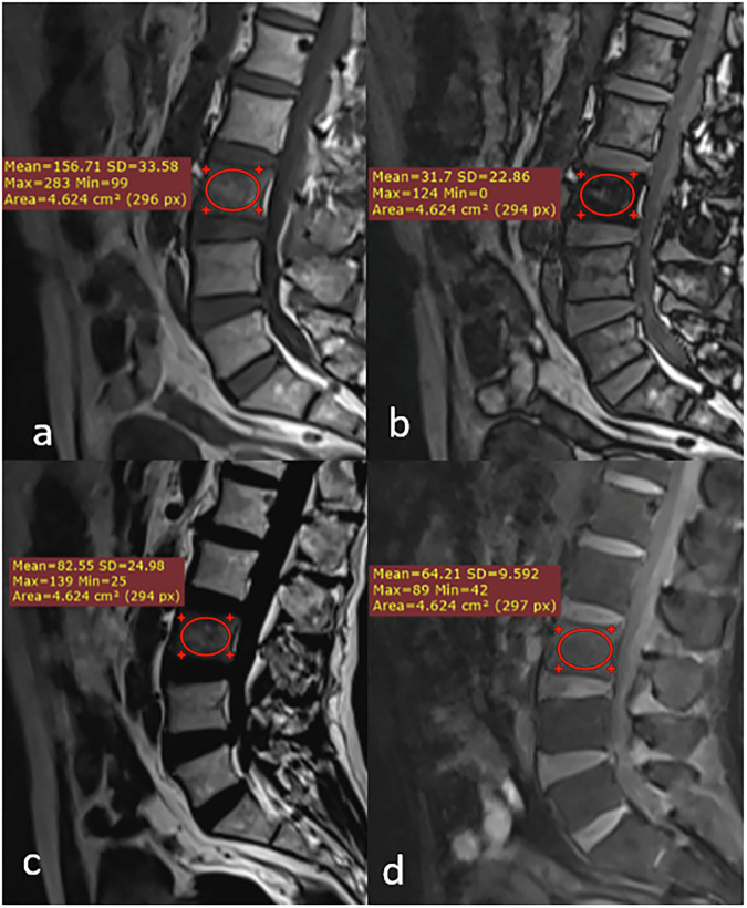
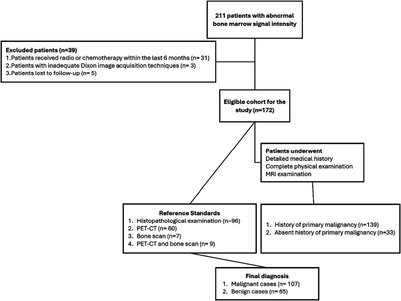
Materials and methods: In this prospective two-center study, 172 patients with BM signal abnormalities underwent standardized 1.5-T MRI protocols, including Dixon sequences. Two musculoskeletal radiologists independently evaluated images and performed quantitative measurements of %drop and %fat. Final diagnoses were established through histopathology (n = 96) or imaging follow-up (n = 76). Diagnostic value was assessed using area under the receiver operating characteristic curve (AUROC), inter-reader reliability using Cohen's κ coefficient.
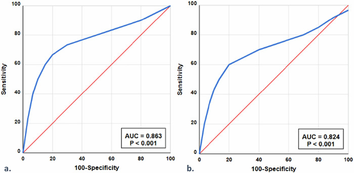
Results: The consensus optimal cutoff was for %drop ≤ 19.8%, yielding 87.2% accuracy, 95.3% sensitivity, and 73.8% specificity, and that for %fat was ≤ 18.3%, achieving 86.6% accuracy, 96.3% sensitivity, and 70.8% specificity. Both metrics showed high diagnostic performance (AUROC 0.824-0.863) and excellent inter-reader reliability (κ > 0.93, p < 0.001). Multivariate analysis identified %drop ≤ 19.8% and %fat ≤ 18.3% as the strongest independent predictors of malignancy, with odds ratio (OR) being 9.38 and 8.85, respectively (p < 0.001). Signal characteristics on Dixon sequences provided additional diagnostic value, with signal voids on fat-only images (OR 7.14) and high signals on water-only images (OR 5.46).
Conclusion: Quantitative MRI Dixon imaging parameters demonstrated high diagnostic accuracy and excellent inter-reader reliability in differentiating benign and malignant BM lesions, supporting their implementation in clinical practice protocols as a reproducible adjunct to conventional MRI.
Relevance statement: Quantitative Dixon MRI provides reproducible, noninvasive differentiation of bone marrow lesions with high diagnostic accuracy across anatomical sites, enhancing clinical decision-making with standardized thresholds while demonstrating excellent inter-center consistency.
Key points: Quantitative Dixon MRI thresholds of %drop ≤ 19.8% and %fat ≤ 18.3% were established as reliable predictors of malignancy in bone marrow lesions. Dixon metrics demonstrated superior diagnostic accuracy (86.6-87.2%), compared to conventional T1-weighted sequences (79.2%). Excellent inter-reader reliability (κ = 0.895-0.943) supports the reproducibility of quantitative Dixon MRI in clinical practice.

 求助内容:
求助内容: 应助结果提醒方式:


