Increased Tc-99m MDP Activities in Multifocal Atypical Ossifying Fibromyxoid Tumor.
IF 0.5
Q4 RADIOLOGY, NUCLEAR MEDICINE & MEDICAL IMAGING
Indian Journal of Nuclear Medicine
Pub Date : 2025-05-01
Epub Date: 2025-08-07
DOI:10.4103/ijnm.ijnm_28_25
引用次数: 0
Abstract
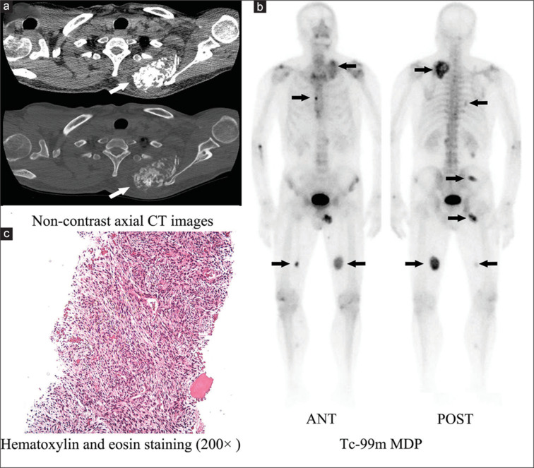
This case demonstrates extraosseous technetium-99m methylene diphosphonate (Tc-99m MDP) accumulation from an atypical ossifying fibromyxoid tumor (OFMT). A 63-year-old man presented with a 10-year history of a gradually enlarging, painless back mass. Physical examination revealed multiple hard, nontender subcutaneous nodules without signs of inflammation or pigmentation. Computed tomography scan of the back showed calcified masses. Whole-body bone scintigraphy revealed areas of increased Tc-99m MDP uptake in subcutaneous regions, suggesting extraosseous uptake. Histopathological examination of the back mass confirmed the diagnosis of atypical OFMT.
Tc-99m MDP在多灶非典型骨化纤维黏液样瘤中的活性升高。
本病例显示非典型骨化纤维黏液样肿瘤(OFMT)的骨外锝-99m二膦酸亚甲基(Tc-99m MDP)积累。男性,63岁,有10年逐渐增大的无痛性背部肿块病史。体格检查显示多发坚硬、无压痛的皮下结节,无炎症或色素沉着迹象。背部计算机断层扫描显示钙化肿块。全身骨显像显示皮下区域Tc-99m MDP摄取增加,提示骨外摄取。背部肿块的组织病理学检查证实了非典型OFMT的诊断。
本文章由计算机程序翻译,如有差异,请以英文原文为准。
求助全文
约1分钟内获得全文
求助全文
来源期刊

Indian Journal of Nuclear Medicine
RADIOLOGY, NUCLEAR MEDICINE & MEDICAL IMAGING-
CiteScore
0.70
自引率
0.00%
发文量
46
 求助内容:
求助内容: 应助结果提醒方式:


