Ozlem Barut Selver, Emil Ahmadli, Muhammed Dara Tas, Banu Yaman, Naim Ceylan, Mozhgan Rezaei Kanavi
{"title":"Orbital Presentation of Eosinophilic Granulomatosis with Polyangiitis: An Interventional Case Report and Literature Review.","authors":"Ozlem Barut Selver, Emil Ahmadli, Muhammed Dara Tas, Banu Yaman, Naim Ceylan, Mozhgan Rezaei Kanavi","doi":"10.18502/jovr.v20.16399","DOIUrl":null,"url":null,"abstract":"<p><strong>Purpose: </strong>To report a case of eosinophilic granulomatosis with polyangiitis (EGPA) initially presenting as orbital involvement, describe its successful management, and provide a comprehensive literature review.</p><p><strong>Case report: </strong>A 33-year-old female patient presented with swelling, redness, tenderness, and a mass under the left upper eyelid for one month. Upper lid eversion showed a multilobulated lesion in the subconjunctival area of the same region. The patient's medical history included asthma and atrial septal defect surgery. Orbital MRI revealed a soft tissue mass infiltrating the superior and lateral aspects of the conal and extraconal regions in the anterior orbit, with extension toward the preseptal area. The lesion underwent incisional biopsy, and histopathological findings were consistent with the diagnosis of EGPA. The patient's blood tests revealed eosinophilia and a negative antineutrophil cytoplasmic antibody. After excluding other similar pathologies such as granulomatosis with polyangiitis, we observed a dramatic regression in her orbital lesion following systemic steroid therapy.</p><p><strong>Conclusion: </strong>The diagnosis of EGPA, a rare clinical presentation, is crucial for ophthalmologists because it provides early recognition of the systemic disease and can help slow its progression by initiating appropriate treatment in a timely manner.</p>","PeriodicalId":16586,"journal":{"name":"Journal of Ophthalmic & Vision Research","volume":"20 ","pages":""},"PeriodicalIF":1.5000,"publicationDate":"2025-08-27","publicationTypes":"Journal Article","fieldsOfStudy":null,"isOpenAccess":false,"openAccessPdf":"https://www.ncbi.nlm.nih.gov/pmc/articles/PMC12396090/pdf/","citationCount":"0","resultStr":null,"platform":"Semanticscholar","paperid":null,"PeriodicalName":"Journal of Ophthalmic & Vision Research","FirstCategoryId":"1085","ListUrlMain":"https://doi.org/10.18502/jovr.v20.16399","RegionNum":0,"RegionCategory":null,"ArticlePicture":[],"TitleCN":null,"AbstractTextCN":null,"PMCID":null,"EPubDate":"2025/1/1 0:00:00","PubModel":"eCollection","JCR":"Q3","JCRName":"OPHTHALMOLOGY","Score":null,"Total":0}
引用次数: 0
Abstract
Purpose: To report a case of eosinophilic granulomatosis with polyangiitis (EGPA) initially presenting as orbital involvement, describe its successful management, and provide a comprehensive literature review.
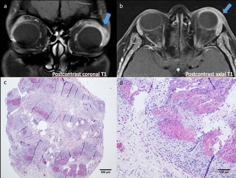
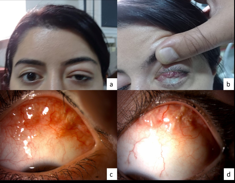
Case report: A 33-year-old female patient presented with swelling, redness, tenderness, and a mass under the left upper eyelid for one month. Upper lid eversion showed a multilobulated lesion in the subconjunctival area of the same region. The patient's medical history included asthma and atrial septal defect surgery. Orbital MRI revealed a soft tissue mass infiltrating the superior and lateral aspects of the conal and extraconal regions in the anterior orbit, with extension toward the preseptal area. The lesion underwent incisional biopsy, and histopathological findings were consistent with the diagnosis of EGPA. The patient's blood tests revealed eosinophilia and a negative antineutrophil cytoplasmic antibody. After excluding other similar pathologies such as granulomatosis with polyangiitis, we observed a dramatic regression in her orbital lesion following systemic steroid therapy.
Conclusion: The diagnosis of EGPA, a rare clinical presentation, is crucial for ophthalmologists because it provides early recognition of the systemic disease and can help slow its progression by initiating appropriate treatment in a timely manner.

 求助内容:
求助内容: 应助结果提醒方式:


