Stefanie Bauer, Karina Hofmann, Miriam Weiss, Christian Musahl, Lukas Andereggen, Gerrit A Schubert
{"title":"Targeted decompression to address long-standing anatomical distortion in a case of extensive, bilateral en plaque meningiomas: illustrative case.","authors":"Stefanie Bauer, Karina Hofmann, Miriam Weiss, Christian Musahl, Lukas Andereggen, Gerrit A Schubert","doi":"10.3171/CASE25382","DOIUrl":null,"url":null,"abstract":"<p><strong>Background: </strong>Meningioma en plaque (MEP) is a rare subtype of meningioma with a carpet-like growth pattern, often causing hyperostosis. Even rarer is the presentation of bilateral MEP posing diagnostic and therapeutic challenges. Management of MEP usually entails early complete resection. Follow-up may be considered for asymptomatic and slow-growing tumors.</p><p><strong>Observations: </strong>The authors present the case of a 52-year-old patient with progressive optic neuropathy due to bilateral hyperostotic frontotemporal MEP. Long-standing intracranial hypertension caused by mass effect had led to formation of a suprachiasmatic cyst deflecting the optic chiasm. The authors aimed to lower the intracranial pressure indirectly by performing a targeted, right-sided craniectomy with tumor removal and mesh cranioplasty. Postoperatively, reconstitution of brain anatomy and stabilization of the optic neuropathy were observed.</p><p><strong>Lessons: </strong>To the authors' knowledge, this is the first case report addressing targeted decompression for severe, long-standing anatomical distortion due to bilateral hyperostotic MEP. Complete resection as recommended for MEPs would have been associated with a high risk of perioperative morbidity in this case. In case of neurological deterioration due to decompensated intracranial hypertension, indirect decompression can effectively address chronic distortion of anatomy. This case report highlights the need for individualized management strategies for extensive MEPs. https://thejns.org/doi/10.3171/CASE25382.</p>","PeriodicalId":94098,"journal":{"name":"Journal of neurosurgery. Case lessons","volume":"10 10","pages":""},"PeriodicalIF":0.0000,"publicationDate":"2025-09-08","publicationTypes":"Journal Article","fieldsOfStudy":null,"isOpenAccess":false,"openAccessPdf":"https://www.ncbi.nlm.nih.gov/pmc/articles/PMC12416326/pdf/","citationCount":"0","resultStr":null,"platform":"Semanticscholar","paperid":null,"PeriodicalName":"Journal of neurosurgery. Case lessons","FirstCategoryId":"1085","ListUrlMain":"https://doi.org/10.3171/CASE25382","RegionNum":0,"RegionCategory":null,"ArticlePicture":[],"TitleCN":null,"AbstractTextCN":null,"PMCID":null,"EPubDate":"","PubModel":"","JCR":"","JCRName":"","Score":null,"Total":0}
引用次数: 0
Abstract
Background: Meningioma en plaque (MEP) is a rare subtype of meningioma with a carpet-like growth pattern, often causing hyperostosis. Even rarer is the presentation of bilateral MEP posing diagnostic and therapeutic challenges. Management of MEP usually entails early complete resection. Follow-up may be considered for asymptomatic and slow-growing tumors.
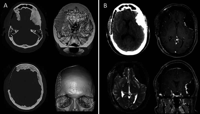
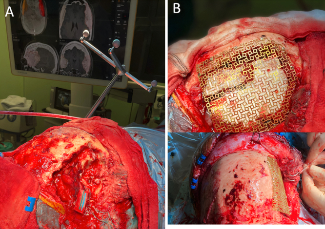
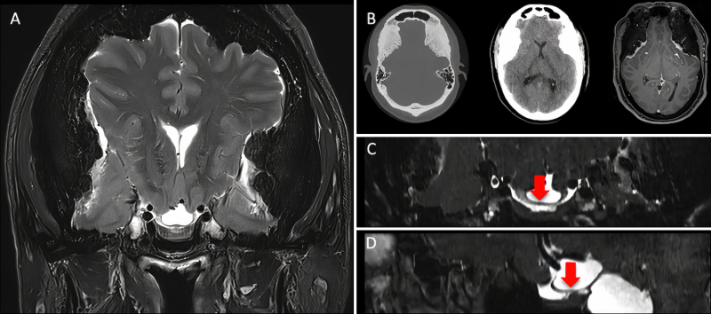
Observations: The authors present the case of a 52-year-old patient with progressive optic neuropathy due to bilateral hyperostotic frontotemporal MEP. Long-standing intracranial hypertension caused by mass effect had led to formation of a suprachiasmatic cyst deflecting the optic chiasm. The authors aimed to lower the intracranial pressure indirectly by performing a targeted, right-sided craniectomy with tumor removal and mesh cranioplasty. Postoperatively, reconstitution of brain anatomy and stabilization of the optic neuropathy were observed.
Lessons: To the authors' knowledge, this is the first case report addressing targeted decompression for severe, long-standing anatomical distortion due to bilateral hyperostotic MEP. Complete resection as recommended for MEPs would have been associated with a high risk of perioperative morbidity in this case. In case of neurological deterioration due to decompensated intracranial hypertension, indirect decompression can effectively address chronic distortion of anatomy. This case report highlights the need for individualized management strategies for extensive MEPs. https://thejns.org/doi/10.3171/CASE25382.

 求助内容:
求助内容: 应助结果提醒方式:


