Sara Smeets, Thomas Daenekindt, Frank Weyns, Christophe Oosterbos
{"title":"CPPD-related basilar impression with vertebral artery dissection and subsequent subarachnoid hemorrhage: illustrative case.","authors":"Sara Smeets, Thomas Daenekindt, Frank Weyns, Christophe Oosterbos","doi":"10.3171/CASE25332","DOIUrl":null,"url":null,"abstract":"<p><strong>Background: </strong>Calcium pyrophosphate dihydrate (CPPD) deposition disease at the craniocervical junction (CCJ) typically presents with a retro-odontoid pseudotumor. Here, the authors report a case of CPPD-induced basilar impression, causing vertebral artery (VA) dissection and hemorrhage.</p><p><strong>Observations: </strong>A 65-year-old male presented with worsening chronic cervicalgia, occipital headaches, and unstable tandem gait. CT revealed basilar impression with severe atlantoaxial erosion and instability, absent on imaging 8 months prior. MRI showed high-grade CCJ stenosis without myelopathy, due to CPPD. Before treatment could be initiated, the patient was suddenly found unresponsive. CT showed posterior fossa hemorrhage due to VA dissection, with secondary obstructive hydrocephalus. An external ventricular drain was placed, followed by coil VA embolization. After halo vest placement, CCJ alignment was restored and the patient underwent occipitocervical fusion. Following prolonged rehabilitation, he regained independent living without significant neurological sequelae.</p><p><strong>Lessons: </strong>This is the first reported case of VA dissection with posterior fossa hemorrhage secondary to basilar impression due to CPPD. Inflammatory arthritides of the CCJ may cause rapidly progressive atlantoaxial erosion, predisposing one to unstable basilar impression and VA dissection. Awareness of this risk is crucial in the surgical management of inflammatory basilar impression, where preoperative vascular imaging may be warranted in selected cases. https://thejns.org/doi/10.3171/CASE25332.</p>","PeriodicalId":94098,"journal":{"name":"Journal of neurosurgery. Case lessons","volume":"10 10","pages":""},"PeriodicalIF":0.0000,"publicationDate":"2025-09-08","publicationTypes":"Journal Article","fieldsOfStudy":null,"isOpenAccess":false,"openAccessPdf":"https://www.ncbi.nlm.nih.gov/pmc/articles/PMC12416330/pdf/","citationCount":"0","resultStr":null,"platform":"Semanticscholar","paperid":null,"PeriodicalName":"Journal of neurosurgery. Case lessons","FirstCategoryId":"1085","ListUrlMain":"https://doi.org/10.3171/CASE25332","RegionNum":0,"RegionCategory":null,"ArticlePicture":[],"TitleCN":null,"AbstractTextCN":null,"PMCID":null,"EPubDate":"","PubModel":"","JCR":"","JCRName":"","Score":null,"Total":0}
引用次数: 0
Abstract
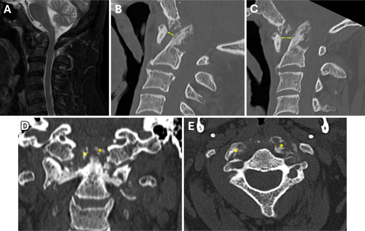
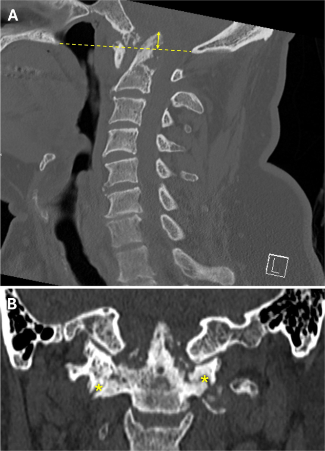
Background: Calcium pyrophosphate dihydrate (CPPD) deposition disease at the craniocervical junction (CCJ) typically presents with a retro-odontoid pseudotumor. Here, the authors report a case of CPPD-induced basilar impression, causing vertebral artery (VA) dissection and hemorrhage.
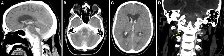
Observations: A 65-year-old male presented with worsening chronic cervicalgia, occipital headaches, and unstable tandem gait. CT revealed basilar impression with severe atlantoaxial erosion and instability, absent on imaging 8 months prior. MRI showed high-grade CCJ stenosis without myelopathy, due to CPPD. Before treatment could be initiated, the patient was suddenly found unresponsive. CT showed posterior fossa hemorrhage due to VA dissection, with secondary obstructive hydrocephalus. An external ventricular drain was placed, followed by coil VA embolization. After halo vest placement, CCJ alignment was restored and the patient underwent occipitocervical fusion. Following prolonged rehabilitation, he regained independent living without significant neurological sequelae.
Lessons: This is the first reported case of VA dissection with posterior fossa hemorrhage secondary to basilar impression due to CPPD. Inflammatory arthritides of the CCJ may cause rapidly progressive atlantoaxial erosion, predisposing one to unstable basilar impression and VA dissection. Awareness of this risk is crucial in the surgical management of inflammatory basilar impression, where preoperative vascular imaging may be warranted in selected cases. https://thejns.org/doi/10.3171/CASE25332.

 求助内容:
求助内容: 应助结果提醒方式:


