Franco Rubino, Peter Harris, Abir L Mukherjee, Walter C Jean
{"title":"High-grade astrocytoma with piloid features resected with an exoscopic supracerebellar infratentorial approach: illustrative case.","authors":"Franco Rubino, Peter Harris, Abir L Mukherjee, Walter C Jean","doi":"10.3171/CASE25390","DOIUrl":null,"url":null,"abstract":"<p><strong>Background: </strong>High-grade astrocytoma with piloid features (HGAP) was recently added to the WHO 2021 CNS classification system among the group of circumscribed astrocytic gliomas. These tumors present with high-grade piloid histology with similarities to glioblastoma. HGAPs in the pineal region become particularly challenging due to its deep location and proximity to deep venous structures, the midbrain, and the thalamus. Herein, the authors present the case of a patient with an HGAP located in the pineal region. The tumor was resected using an exoscopic supracerebellar/infratentorial approach.</p><p><strong>Observations: </strong>A 69-year-old man presented with a pineal gland mass causing obstructive hydrocephalus. Resection using an exoscope revealed an HGAP. Postoperatively, he developed posterior fossa syndrome but was discharged without neurological deficits after 30 days.</p><p><strong>Lessons: </strong>HGAP, a distinct glioma subtype identified in 2021, presents a mix of low- and high-grade features. It shares histological traits with pilocytic astrocytoma and glioblastoma, requiring DNA methylation profiling for diagnosis. Radiologically, these lesions have a T2-FLAIR mismatch and uneven post-gadolinium enhancement. Treatment is still uncertain, although adjuvant chemoradiation therapy with temozolomide may be used. The prognosis is poor, with a 5-year survival rate of approximately 50%. For pineal region locations, the exoscope offers enhanced magnification, depth perception, and ergonomic benefits, improving surgical precision. https://thejns.org/doi/10.3171/CASE25390.</p>","PeriodicalId":94098,"journal":{"name":"Journal of neurosurgery. Case lessons","volume":"10 10","pages":""},"PeriodicalIF":0.0000,"publicationDate":"2025-09-08","publicationTypes":"Journal Article","fieldsOfStudy":null,"isOpenAccess":false,"openAccessPdf":"https://www.ncbi.nlm.nih.gov/pmc/articles/PMC12416328/pdf/","citationCount":"0","resultStr":null,"platform":"Semanticscholar","paperid":null,"PeriodicalName":"Journal of neurosurgery. Case lessons","FirstCategoryId":"1085","ListUrlMain":"https://doi.org/10.3171/CASE25390","RegionNum":0,"RegionCategory":null,"ArticlePicture":[],"TitleCN":null,"AbstractTextCN":null,"PMCID":null,"EPubDate":"","PubModel":"","JCR":"","JCRName":"","Score":null,"Total":0}
引用次数: 0
Abstract
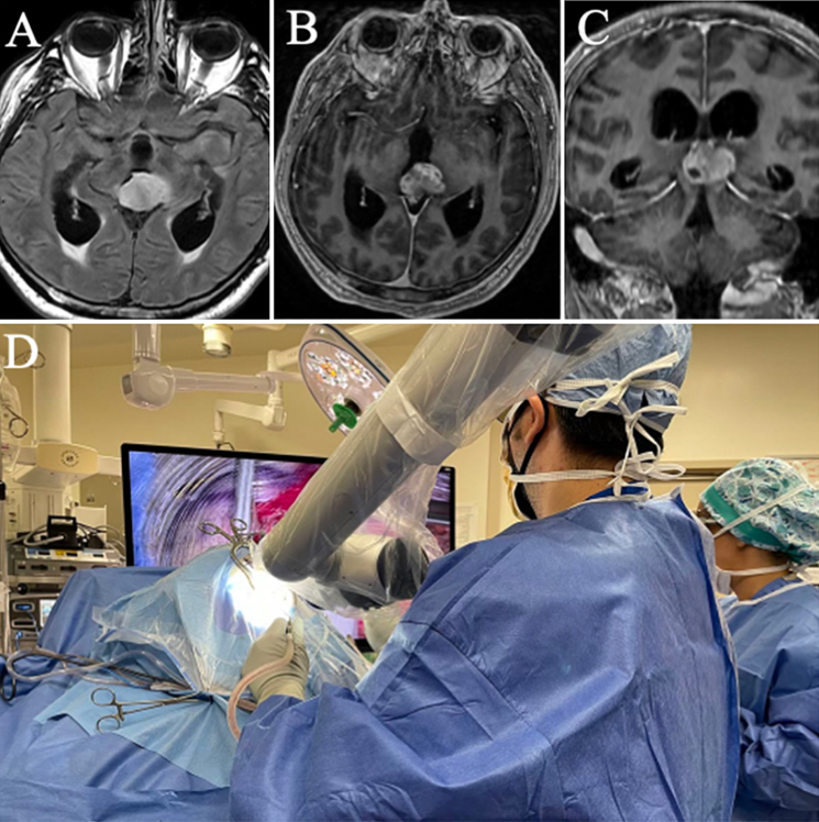
Background: High-grade astrocytoma with piloid features (HGAP) was recently added to the WHO 2021 CNS classification system among the group of circumscribed astrocytic gliomas. These tumors present with high-grade piloid histology with similarities to glioblastoma. HGAPs in the pineal region become particularly challenging due to its deep location and proximity to deep venous structures, the midbrain, and the thalamus. Herein, the authors present the case of a patient with an HGAP located in the pineal region. The tumor was resected using an exoscopic supracerebellar/infratentorial approach.
Observations: A 69-year-old man presented with a pineal gland mass causing obstructive hydrocephalus. Resection using an exoscope revealed an HGAP. Postoperatively, he developed posterior fossa syndrome but was discharged without neurological deficits after 30 days.
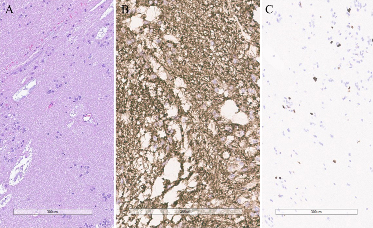
Lessons: HGAP, a distinct glioma subtype identified in 2021, presents a mix of low- and high-grade features. It shares histological traits with pilocytic astrocytoma and glioblastoma, requiring DNA methylation profiling for diagnosis. Radiologically, these lesions have a T2-FLAIR mismatch and uneven post-gadolinium enhancement. Treatment is still uncertain, although adjuvant chemoradiation therapy with temozolomide may be used. The prognosis is poor, with a 5-year survival rate of approximately 50%. For pineal region locations, the exoscope offers enhanced magnification, depth perception, and ergonomic benefits, improving surgical precision. https://thejns.org/doi/10.3171/CASE25390.


 求助内容:
求助内容: 应助结果提醒方式:


