{"title":"Evaluation of Cartilage Degeneration Using MRI T1ρ Mapping after Locomotion Training in Patients with Early Knee Osteoarthritis.","authors":"Yuji Arai, Kazuteru Ryu, Shuji Nakagawa, Koichi Idei, Atsuo Inoue, Kenji Takahashi","doi":"10.2490/prm.20250020","DOIUrl":null,"url":null,"abstract":"<p><strong>Objectives: </strong>: This study aimed to assess the qualitative effects of locomotion training (LT) on articular cartilage using magnetic resonance imaging T1ρ mapping.</p><p><strong>Methods: </strong>: Fifteen patients with early knee osteoarthritis participated in the study. They performed a series of exercises, including one-leg stands, squats, heel raises, and front lunges, on a daily basis for 12 weeks. Knee joint function and physical performance were evaluated before and 3 months after completing the LT program. Additionally, a questionnaire was administered to assess patient-reported outcomes. The quality of articular cartilage was evaluated through magnetic resonance imaging using T1ρ mapping. T1ρ values were quantified on sagittal planes with ten regions of interest.</p><p><strong>Results: </strong>: Regarding physical function, the time required for one-leg stands was significantly increased following LT, and the right functional reach test and the 30-s chair stand test showed improvement. T1ρ values were significantly reduced at 0° relative to the anatomical axis of the femur, whereas other regions of interest showed no significant change after LT.</p><p><strong>Conclusions: </strong>: LT significantly improved muscle strength and balance in patients with early knee osteoarthritis. LT also improved the T1ρ values of articular cartilage at 0° relative to the anatomical axis of the femur. This change may reflect that LT mitigates cartilage degeneration following the application of moderate mechanical stress to the loading region. We propose that LT represents a safe and effective exercise therapy for early knee osteoarthritis, with the potential to improve both motor function and articular cartilage quality.</p>","PeriodicalId":74584,"journal":{"name":"Progress in rehabilitation medicine","volume":"10 ","pages":"20250020"},"PeriodicalIF":1.5000,"publicationDate":"2025-09-05","publicationTypes":"Journal Article","fieldsOfStudy":null,"isOpenAccess":false,"openAccessPdf":"https://www.ncbi.nlm.nih.gov/pmc/articles/PMC12408120/pdf/","citationCount":"0","resultStr":null,"platform":"Semanticscholar","paperid":null,"PeriodicalName":"Progress in rehabilitation medicine","FirstCategoryId":"1085","ListUrlMain":"https://doi.org/10.2490/prm.20250020","RegionNum":0,"RegionCategory":null,"ArticlePicture":[],"TitleCN":null,"AbstractTextCN":null,"PMCID":null,"EPubDate":"2025/1/1 0:00:00","PubModel":"eCollection","JCR":"","JCRName":"","Score":null,"Total":0}
引用次数: 0
Abstract
Objectives: : This study aimed to assess the qualitative effects of locomotion training (LT) on articular cartilage using magnetic resonance imaging T1ρ mapping.
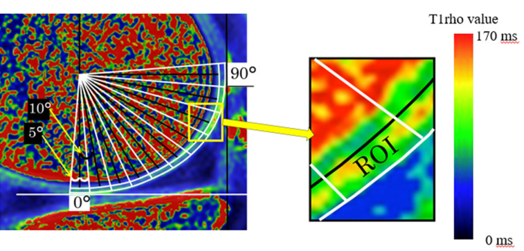
Methods: : Fifteen patients with early knee osteoarthritis participated in the study. They performed a series of exercises, including one-leg stands, squats, heel raises, and front lunges, on a daily basis for 12 weeks. Knee joint function and physical performance were evaluated before and 3 months after completing the LT program. Additionally, a questionnaire was administered to assess patient-reported outcomes. The quality of articular cartilage was evaluated through magnetic resonance imaging using T1ρ mapping. T1ρ values were quantified on sagittal planes with ten regions of interest.
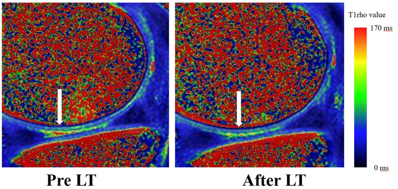
Results: : Regarding physical function, the time required for one-leg stands was significantly increased following LT, and the right functional reach test and the 30-s chair stand test showed improvement. T1ρ values were significantly reduced at 0° relative to the anatomical axis of the femur, whereas other regions of interest showed no significant change after LT.
Conclusions: : LT significantly improved muscle strength and balance in patients with early knee osteoarthritis. LT also improved the T1ρ values of articular cartilage at 0° relative to the anatomical axis of the femur. This change may reflect that LT mitigates cartilage degeneration following the application of moderate mechanical stress to the loading region. We propose that LT represents a safe and effective exercise therapy for early knee osteoarthritis, with the potential to improve both motor function and articular cartilage quality.

 求助内容:
求助内容: 应助结果提醒方式:


