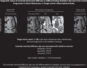
Diagnostic value of centrally restricted diffusion in differentiating radiation necrosis from tumor progression in brain metastases: A single-center observational study
IF 3.3
3区 医学
Q2 CLINICAL NEUROLOGY
引用次数: 0
Abstract
Background
Distinguishing radiation necrosis (RN) from true progression (TP) in irradiated brain metastases is challenging. We evaluated the diagnostic performance of the centrally restricted diffusion sign on diffusion-weighted imaging (DWI).
Methods
From August 2014 to August 2024, we screened 321 patients with histologically confirmed brain metastases treated with radiation therapy and follow-up MRI for new or enlarging necrotic lesions ≥1 cm. Two board-certified neuroradiologists independently assessed the centrally restricted diffusion sign—central hyperintensity on b1000 images with corresponding ADC reduction—by rigidly co-registering DWI to postcontrast 3D T1-weighted sequences. Quantitative analysis included mean ADC measurement within manually drawn regions of interest in the necrotic core and the contrast-enhancing rim. Final diagnoses were established by histopathology (n = 17) or multidisciplinary consensus (n = 90).
Results
Of 107 patients (median age, 62 years; 57.9 % male), 62 had TP and 45 had RN. Median interval from radiotherapy completion to index MRI was 10.8 months. Overall survival was longer in patients with RN (median not reached) than in those with TP (17.5 months; P < 0.0001). Interobserver agreement for the centrally restricted diffusion sign was moderate (κ =0.55). The sign appeared in 34/45 RN cases and 13/62 TP cases (P < 0.0001). For RN diagnosis, sensitivity was 75.6 %, specificity 79.0 %, and accuracy 77.6 %. Quantitative ADC metrics did not enhance performance.
Conclusion
The centrally restricted diffusion sign on DWI may aid differentiation of RN from TP in irradiated brain metastases, despite moderate interrater reliability.
中枢限制性弥散在鉴别脑转移灶放射性坏死与肿瘤进展中的诊断价值:一项单中心观察研究。
背景:区分放射性脑转移瘤的放射性坏死(RN)和真进展(TP)是具有挑战性的。我们评估了弥散加权成像(DWI)上中央限制性弥散征象的诊断性能。方法:从2014年8月至2024年8月,我们筛选了321例组织学证实的脑转移患者,这些患者接受了放疗,并随访MRI,发现新的或扩大的坏死灶≥1 cm。两名委员会认证的神经放射学家通过严格地将DWI与对比后的3D t1加权序列共同配准,独立评估了b1000图像上的中心限制性弥散征象-相应ADC降低的中心高强度。定量分析包括在坏死核心和对比度增强边缘手动绘制的感兴趣区域内的平均ADC测量。最终诊断由组织病理学(n=17)或多学科共识(n=90)确定。结果:107例患者中位年龄62岁,男性57.9%,TP 62例,RN 45例。从放疗完成到MRI指数的中位时间间隔为10.8个月。结论:DWI上的中枢性弥散受限征象可能有助于放射脑转移灶中RN与TP的鉴别,尽管两者间的可靠性不高。
本文章由计算机程序翻译,如有差异,请以英文原文为准。
求助全文
约1分钟内获得全文
求助全文
来源期刊
Journal of Neuroradiology
医学-核医学
CiteScore
6.10
自引率
5.70%
发文量
142
审稿时长
6-12 weeks
期刊介绍:
The Journal of Neuroradiology is a peer-reviewed journal, publishing worldwide clinical and basic research in the field of diagnostic and Interventional neuroradiology, translational and molecular neuroimaging, and artificial intelligence in neuroradiology.
The Journal of Neuroradiology considers for publication articles, reviews, technical notes and letters to the editors (correspondence section), provided that the methodology and scientific content are of high quality, and that the results will have substantial clinical impact and/or physiological importance.
 求助内容:
求助内容: 应助结果提醒方式:


