{"title":"Lumbosacral intradural-extramedullary intervertebral disc extrusion in a cat.","authors":"Lauryn Cooper, Marc Kent","doi":"10.1177/20551169251352386","DOIUrl":null,"url":null,"abstract":"<p><strong>Case summary: </strong>A 10-year-old female spayed domestic shorthair cat was evaluated for a 6-week history of abnormal tail carriage and constipation. Examination revealed tail paresis and pain over the lumbosacral and sacrocaudal articulations and on tail manipulation. MRI revealed a contrast-enhancing mass within the vertebral canal over the lumbosacral disc space, compressing the cauda equina. The mass filled the epidural space, resulting in complete attenuation of the cerebrospinal fluid signal. Laminectomy and durotomy were performed over L7-S1, revealing white, firm material within the subarachnoid space. Microscopically, the material was consistent with degenerative intervertebral disc material. Postoperatively, the clinical signs resolved completely.</p><p><strong>Relevance and novel information: </strong>Intervertebral disc herniation (IVDH) is uncommon in cats, with most cases involving extradural compression of nervous tissue. Reports describing intramedullary intervertebral disc extrusions in cats are rare. To the authors' knowledge, the present case is the first reported intradural-extramedullary intervertebral disc extrusion in a cat. Although MRI can often delineate extradural lesions, it can be insensitive in differentiating intradural-extramedullary from intramedullary lesions. In the present case, the location of the lesion within the vertebral canal at the lumbosacral disc space made the determination of the lesion's location with respect to the meninges challenging. Moreover, the strong contrast enhancement of the lesion raised an index of suspicion for neoplasia. Surgical intervention and histopathology confirmed an intradural-extramedullary IVDH. The present case adds to a growing body of literature regarding IVDH in cats and details the imaging findings of intradural-extramedullary IVDH in a cat.</p>","PeriodicalId":36588,"journal":{"name":"Journal of Feline Medicine and Surgery Open Reports","volume":"11 2","pages":"20551169251352386"},"PeriodicalIF":0.7000,"publicationDate":"2025-09-04","publicationTypes":"Journal Article","fieldsOfStudy":null,"isOpenAccess":false,"openAccessPdf":"https://www.ncbi.nlm.nih.gov/pmc/articles/PMC12411724/pdf/","citationCount":"0","resultStr":null,"platform":"Semanticscholar","paperid":null,"PeriodicalName":"Journal of Feline Medicine and Surgery Open Reports","FirstCategoryId":"1085","ListUrlMain":"https://doi.org/10.1177/20551169251352386","RegionNum":0,"RegionCategory":null,"ArticlePicture":[],"TitleCN":null,"AbstractTextCN":null,"PMCID":null,"EPubDate":"2025/7/1 0:00:00","PubModel":"eCollection","JCR":"Q3","JCRName":"VETERINARY SCIENCES","Score":null,"Total":0}
引用次数: 0
Abstract
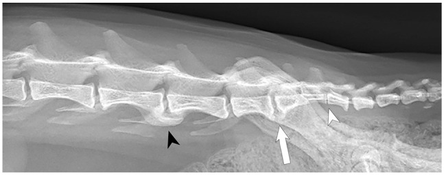
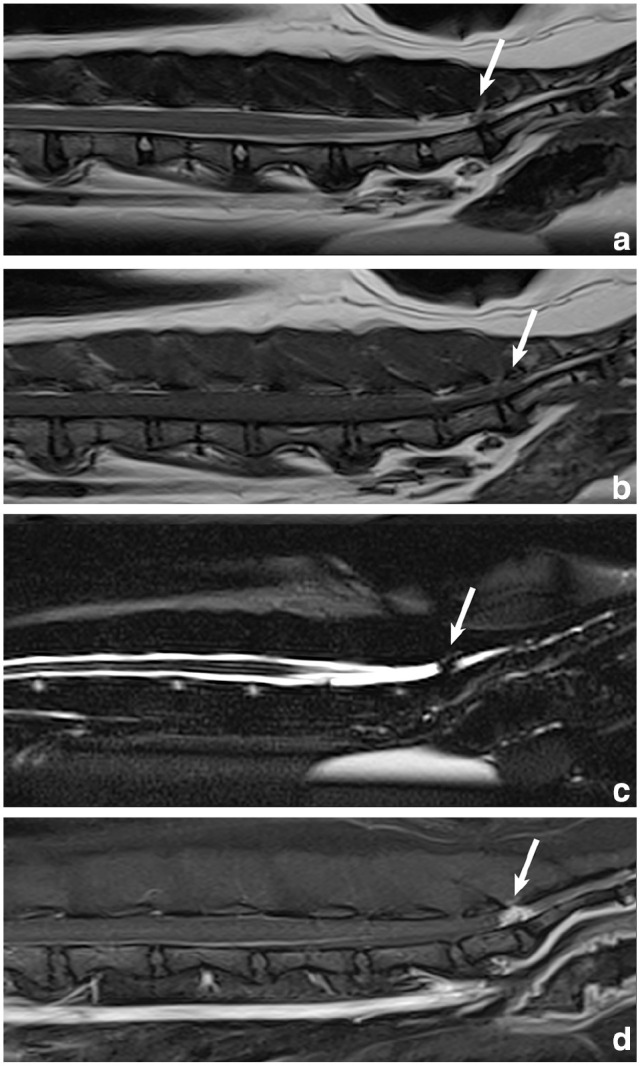
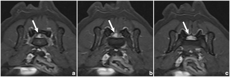
Case summary: A 10-year-old female spayed domestic shorthair cat was evaluated for a 6-week history of abnormal tail carriage and constipation. Examination revealed tail paresis and pain over the lumbosacral and sacrocaudal articulations and on tail manipulation. MRI revealed a contrast-enhancing mass within the vertebral canal over the lumbosacral disc space, compressing the cauda equina. The mass filled the epidural space, resulting in complete attenuation of the cerebrospinal fluid signal. Laminectomy and durotomy were performed over L7-S1, revealing white, firm material within the subarachnoid space. Microscopically, the material was consistent with degenerative intervertebral disc material. Postoperatively, the clinical signs resolved completely.
Relevance and novel information: Intervertebral disc herniation (IVDH) is uncommon in cats, with most cases involving extradural compression of nervous tissue. Reports describing intramedullary intervertebral disc extrusions in cats are rare. To the authors' knowledge, the present case is the first reported intradural-extramedullary intervertebral disc extrusion in a cat. Although MRI can often delineate extradural lesions, it can be insensitive in differentiating intradural-extramedullary from intramedullary lesions. In the present case, the location of the lesion within the vertebral canal at the lumbosacral disc space made the determination of the lesion's location with respect to the meninges challenging. Moreover, the strong contrast enhancement of the lesion raised an index of suspicion for neoplasia. Surgical intervention and histopathology confirmed an intradural-extramedullary IVDH. The present case adds to a growing body of literature regarding IVDH in cats and details the imaging findings of intradural-extramedullary IVDH in a cat.

 求助内容:
求助内容: 应助结果提醒方式:


