Nicola Valsecchi, Elham Sadeghi, Elli Davis, Mohammed Nasar Ibrahim, Nasiq Hasan, Sandeep Chandra Bollepalli, Sumit Randhir Singh, Luigi Fontana, Jose Alain Sahel, Kiran Kumar Vupparaboina, Jay Chhablani
{"title":"Three-Dimensional Choroidal Vessels Assessment in Fellow Eyes of Patients With Central Serous Chorioretinopathy.","authors":"Nicola Valsecchi, Elham Sadeghi, Elli Davis, Mohammed Nasar Ibrahim, Nasiq Hasan, Sandeep Chandra Bollepalli, Sumit Randhir Singh, Luigi Fontana, Jose Alain Sahel, Kiran Kumar Vupparaboina, Jay Chhablani","doi":"10.1167/tvst.14.9.10","DOIUrl":null,"url":null,"abstract":"<p><strong>Purpose: </strong>To evaluate choroidal vasculature using a novel three-dimensional algorithm in fellow eyes of patients with unilateral chronic central serous chorioretinopathy (cCSC).</p><p><strong>Methods: </strong>Patients with unilateral cCSC were retrospectively included. Automated choroidal segmentation was conducted using a deep-learning ResUNet model. Phansalkar thresholding was applied to binarize choroidal vasculature, and three-dimensional maps were created. Mean choroidal vessel diameter, intervessel distance, choroidal thickness, and choroidal vascularity index (CVI) were measured. Linear mixed models were used for statistical analysis.</p><p><strong>Results: </strong>Thirty unilateral cCSC eyes, 22 fellow, and 26 controls were included. Both cCSC and fellow eyes exhibited significant higher mean choroidal vessel diameter compared with controls (362.50 ± 83.23 µm, 276.84 ± 26.89 µm, and 233.28 ± 28.18 µm, respectively; P < 0.001), and in choroidal thickness (288.90 ± 64.77 µm, 269.76 ± 71.17 µm, and 223.97 ± 43.40 µm, respectively; P = 0.001). The intervessel distance was reduced in cCSC and fellow eyes compared with controls (196.53 ± 23.58 µm, 225.05 ± 33.72 µm, and 264.13 ± 46.06 µm, respectively; P < 0.001). Although lower, the CVI was not significantly different in cCSC and fellow eyes compared with controls (38.14 ± 5.55%, 37.23 ± 6.41%, and 40.65 ± 3.53%, respectively; P = 0.066), indicating a possible trend toward a lower CVI.</p><p><strong>Conclusions: </strong>Three-dimensional representation of choroidal vasculature revealed significant changes in both cCSC and fellow eyes, including a larger diameter and reduced spacing compared with healthy controls.</p><p><strong>Translational relevance: </strong>Using a validated deep learning-based three-dimensional method, we observed changes in the choroidal vasculature in both CSC and fellow eyes.</p>","PeriodicalId":23322,"journal":{"name":"Translational Vision Science & Technology","volume":"14 9","pages":"10"},"PeriodicalIF":2.6000,"publicationDate":"2025-09-02","publicationTypes":"Journal Article","fieldsOfStudy":null,"isOpenAccess":false,"openAccessPdf":"https://www.ncbi.nlm.nih.gov/pmc/articles/PMC12422394/pdf/","citationCount":"0","resultStr":null,"platform":"Semanticscholar","paperid":null,"PeriodicalName":"Translational Vision Science & Technology","FirstCategoryId":"3","ListUrlMain":"https://doi.org/10.1167/tvst.14.9.10","RegionNum":3,"RegionCategory":"医学","ArticlePicture":[],"TitleCN":null,"AbstractTextCN":null,"PMCID":null,"EPubDate":"","PubModel":"","JCR":"Q2","JCRName":"OPHTHALMOLOGY","Score":null,"Total":0}
引用次数: 0
Abstract
Purpose: To evaluate choroidal vasculature using a novel three-dimensional algorithm in fellow eyes of patients with unilateral chronic central serous chorioretinopathy (cCSC).
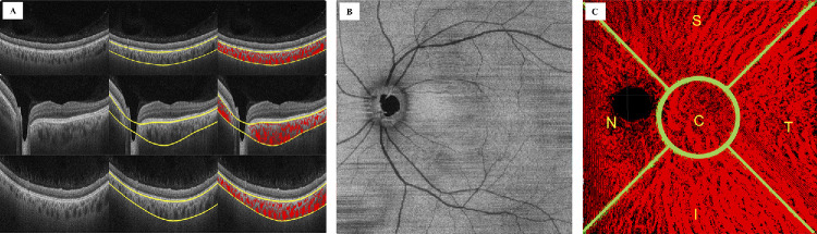
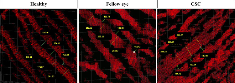
Methods: Patients with unilateral cCSC were retrospectively included. Automated choroidal segmentation was conducted using a deep-learning ResUNet model. Phansalkar thresholding was applied to binarize choroidal vasculature, and three-dimensional maps were created. Mean choroidal vessel diameter, intervessel distance, choroidal thickness, and choroidal vascularity index (CVI) were measured. Linear mixed models were used for statistical analysis.
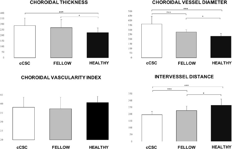
Results: Thirty unilateral cCSC eyes, 22 fellow, and 26 controls were included. Both cCSC and fellow eyes exhibited significant higher mean choroidal vessel diameter compared with controls (362.50 ± 83.23 µm, 276.84 ± 26.89 µm, and 233.28 ± 28.18 µm, respectively; P < 0.001), and in choroidal thickness (288.90 ± 64.77 µm, 269.76 ± 71.17 µm, and 223.97 ± 43.40 µm, respectively; P = 0.001). The intervessel distance was reduced in cCSC and fellow eyes compared with controls (196.53 ± 23.58 µm, 225.05 ± 33.72 µm, and 264.13 ± 46.06 µm, respectively; P < 0.001). Although lower, the CVI was not significantly different in cCSC and fellow eyes compared with controls (38.14 ± 5.55%, 37.23 ± 6.41%, and 40.65 ± 3.53%, respectively; P = 0.066), indicating a possible trend toward a lower CVI.
Conclusions: Three-dimensional representation of choroidal vasculature revealed significant changes in both cCSC and fellow eyes, including a larger diameter and reduced spacing compared with healthy controls.
Translational relevance: Using a validated deep learning-based three-dimensional method, we observed changes in the choroidal vasculature in both CSC and fellow eyes.
目的:利用一种新的三维算法评估单侧慢性中枢性浆液性脉络膜视网膜病变(cCSC)患者的同眼脉络膜血管。方法:回顾性分析单侧cCSC患者。使用深度学习的ResUNet模型进行脉络膜自动分割。应用phansalar阈值法二值化脉络膜血管,并创建三维地图。测量平均脉络膜血管直径、血管间距离、脉络膜厚度和脉络膜血管指数(CVI)。采用线性混合模型进行统计分析。结果:30只单侧cCSC眼,22只患者,26只对照组。cCSC组和其他眼组的平均脉膜血管直径(分别为362.50±83.23µm、276.84±26.89µm和233.28±28.18µm, P < 0.001)和脉膜厚度(分别为288.90±64.77µm、269.76±71.17µm和223.97±43.40µm, P = 0.001)均显著高于对照组。与对照组相比,cCSC和其他眼的血管间距离缩短(分别为196.53±23.58µm, 225.05±33.72µm和264.13±46.06µm, P < 0.001)。虽然CVI较低,但cCSC和其他眼的CVI与对照组相比差异不显著(分别为38.14±5.55%,37.23±6.41%和40.65±3.53%,P = 0.066),表明CVI可能有降低的趋势。结论:脉络膜血管的三维表征显示cCSC和其他眼的明显变化,包括与健康对照相比直径更大,间距更小。翻译相关性:使用经过验证的基于深度学习的三维方法,我们观察了CSC和其他眼睛的脉络膜血管系统的变化。
期刊介绍:
Translational Vision Science & Technology (TVST), an official journal of the Association for Research in Vision and Ophthalmology (ARVO), an international organization whose purpose is to advance research worldwide into understanding the visual system and preventing, treating and curing its disorders, is an online, open access, peer-reviewed journal emphasizing multidisciplinary research that bridges the gap between basic research and clinical care. A highly qualified and diverse group of Associate Editors and Editorial Board Members is led by Editor-in-Chief Marco Zarbin, MD, PhD, FARVO.
The journal covers a broad spectrum of work, including but not limited to:
Applications of stem cell technology for regenerative medicine,
Development of new animal models of human diseases,
Tissue bioengineering,
Chemical engineering to improve virus-based gene delivery,
Nanotechnology for drug delivery,
Design and synthesis of artificial extracellular matrices,
Development of a true microsurgical operating environment,
Refining data analysis algorithms to improve in vivo imaging technology,
Results of Phase 1 clinical trials,
Reverse translational ("bedside to bench") research.
TVST seeks manuscripts from scientists and clinicians with diverse backgrounds ranging from basic chemistry to ophthalmic surgery that will advance or change the way we understand and/or treat vision-threatening diseases. TVST encourages the use of color, multimedia, hyperlinks, program code and other digital enhancements.

 求助内容:
求助内容: 应助结果提醒方式:


