{"title":"Comparison of structural brain volume among sedentary normal, overweight and obese adults - A cross-sectional study.","authors":"Nikhil Raj, Rajagopal Kadavigere, Baskaran Chandrasekaran, K Vaishali, Shailesh Nayak, Sneha Ravichandran, Dilip Shettigar, Sathya Sabina Muthu, Cyril Biji, Poovitha Shruthi Paramashiva, Suresh Sukumar","doi":"10.4103/jehp.jehp_1396_24","DOIUrl":null,"url":null,"abstract":"<p><strong>Background: </strong>The relationship between obesity and brain structure remains unclear, particularly in sedentary individuals. This study aimed to compare structural brain volumes among sedentary normal weight, overweight, and obese adults.</p><p><strong>Materials and methods: </strong>In this cross-sectional study, 102 sedentary adults (34 normal weight, 34 overweight, 34 obese) underwent brain MRI scans. Grey matter, white matter, cerebrospinal fluid, and regional brain volumes were measured. Correlations between BMI, physical activity levels, and brain volumes were analyzed within each weight group.</p><p><strong>Results: </strong>No significant differences in overall or regional brain volumes were found between groups. In the normal weight group, BMI positively correlated with right superior temporal gyrus (rSTG) volume (<i>r</i> = 0.358, <i>P</i> < 0.05) and grey matter volume (<i>r</i> = 0.367, <i>P</i> < 0.05). In the obese group, BMI negatively correlated with rSTG volume (<i>r</i> = -0.467, <i>P</i> < 0.01) and positively correlated with self-reported physical activity (<i>r</i> = 0.395, <i>P</i> < 0.05). No significant correlations were observed in the overweight group.</p><p><strong>Conclusions: </strong>This study reveals a complex, non-linear relationship between BMI and brain structure in sedentary adults. The contrasting correlations in normal weight and obese groups suggest potential BMI-related structural changes, particularly in the rSTG. These findings highlight the need for further research on the neurological impacts of obesity in sedentary populations.</p>","PeriodicalId":15581,"journal":{"name":"Journal of Education and Health Promotion","volume":"14 ","pages":"297"},"PeriodicalIF":1.3000,"publicationDate":"2025-07-31","publicationTypes":"Journal Article","fieldsOfStudy":null,"isOpenAccess":false,"openAccessPdf":"https://www.ncbi.nlm.nih.gov/pmc/articles/PMC12413107/pdf/","citationCount":"0","resultStr":null,"platform":"Semanticscholar","paperid":null,"PeriodicalName":"Journal of Education and Health Promotion","FirstCategoryId":"1085","ListUrlMain":"https://doi.org/10.4103/jehp.jehp_1396_24","RegionNum":0,"RegionCategory":null,"ArticlePicture":[],"TitleCN":null,"AbstractTextCN":null,"PMCID":null,"EPubDate":"2025/1/1 0:00:00","PubModel":"eCollection","JCR":"Q3","JCRName":"EDUCATION, SCIENTIFIC DISCIPLINES","Score":null,"Total":0}
引用次数: 0
Abstract
Background: The relationship between obesity and brain structure remains unclear, particularly in sedentary individuals. This study aimed to compare structural brain volumes among sedentary normal weight, overweight, and obese adults.
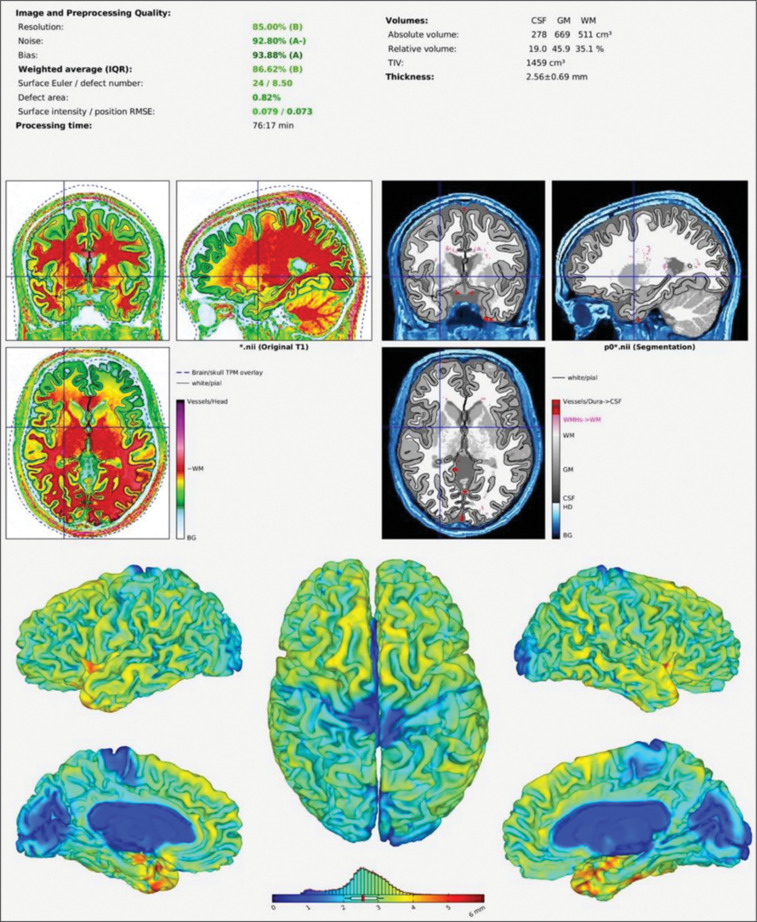
Materials and methods: In this cross-sectional study, 102 sedentary adults (34 normal weight, 34 overweight, 34 obese) underwent brain MRI scans. Grey matter, white matter, cerebrospinal fluid, and regional brain volumes were measured. Correlations between BMI, physical activity levels, and brain volumes were analyzed within each weight group.
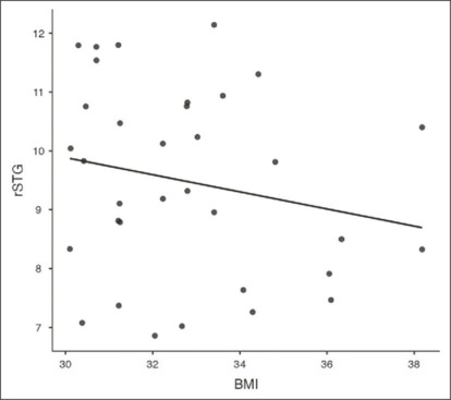
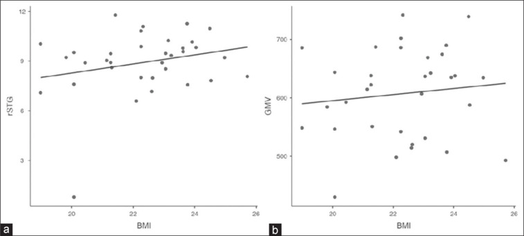
Results: No significant differences in overall or regional brain volumes were found between groups. In the normal weight group, BMI positively correlated with right superior temporal gyrus (rSTG) volume (r = 0.358, P < 0.05) and grey matter volume (r = 0.367, P < 0.05). In the obese group, BMI negatively correlated with rSTG volume (r = -0.467, P < 0.01) and positively correlated with self-reported physical activity (r = 0.395, P < 0.05). No significant correlations were observed in the overweight group.
Conclusions: This study reveals a complex, non-linear relationship between BMI and brain structure in sedentary adults. The contrasting correlations in normal weight and obese groups suggest potential BMI-related structural changes, particularly in the rSTG. These findings highlight the need for further research on the neurological impacts of obesity in sedentary populations.

 求助内容:
求助内容: 应助结果提醒方式:


