Marco Rossi, Claudia Manera, Francesco Saverio Ludovichetti, Christian Bacci
{"title":"Oral Dermoid Cyst in the Mouth Floor: A Case Report and 5-Year Review of the Literature.","authors":"Marco Rossi, Claudia Manera, Francesco Saverio Ludovichetti, Christian Bacci","doi":"10.1155/crid/8851087","DOIUrl":null,"url":null,"abstract":"<p><p><b>Aim:</b> The aim of the study is to present a case of a dermoid cyst in the oral floor of an 80-year-old female patient and discuss its clinical presentation, diagnostic approach, and treatment. <b>Methods:</b> A retrospective case study was conducted at the Department of Maxillofacial Surgery, Aulss 2 Marca Trevigiana. The patient, with a medical history of hypertension, presented with a swelling in the oral floor. A clinical examination was followed by a facial CT scan with and without contrast medium to evaluate the cystic lesion. The patient underwent surgical excision under general anesthesia, with subsequent histopathological analysis. <b>Results:</b> The CT scan revealed a round, homogeneous, hyperdense formation with no enhancement after contrast administration. Surgical excision was performed using an intraoral approach, and histopathology confirmed the diagnosis of a dermoid cyst with an epithelial lining of predominantly squamous type. The patient experienced no complications, and follow-up at 9 days and 2 months showed complete recovery. <b>Conclusion:</b> A dermoid cyst of the oral cavity, though rare in adults, should be considered in the differential diagnosis of oral floor swellings. Early diagnosis through clinical examination and imaging, followed by surgical excision, provides an effective and curative treatment.</p>","PeriodicalId":46841,"journal":{"name":"Case Reports in Dentistry","volume":"2025 ","pages":"8851087"},"PeriodicalIF":0.9000,"publicationDate":"2025-08-30","publicationTypes":"Journal Article","fieldsOfStudy":null,"isOpenAccess":false,"openAccessPdf":"https://www.ncbi.nlm.nih.gov/pmc/articles/PMC12413954/pdf/","citationCount":"0","resultStr":null,"platform":"Semanticscholar","paperid":null,"PeriodicalName":"Case Reports in Dentistry","FirstCategoryId":"1085","ListUrlMain":"https://doi.org/10.1155/crid/8851087","RegionNum":0,"RegionCategory":null,"ArticlePicture":[],"TitleCN":null,"AbstractTextCN":null,"PMCID":null,"EPubDate":"2025/1/1 0:00:00","PubModel":"eCollection","JCR":"Q4","JCRName":"DENTISTRY, ORAL SURGERY & MEDICINE","Score":null,"Total":0}
引用次数: 0
Abstract
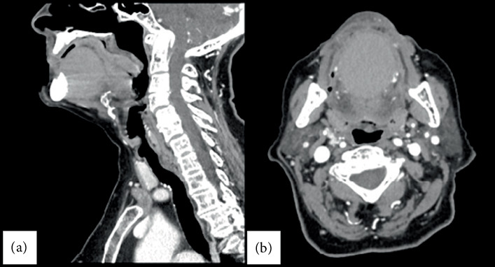
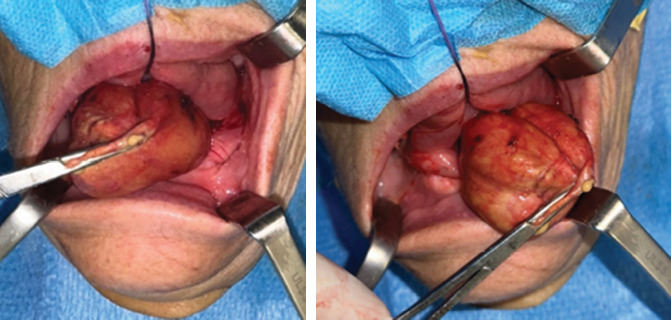
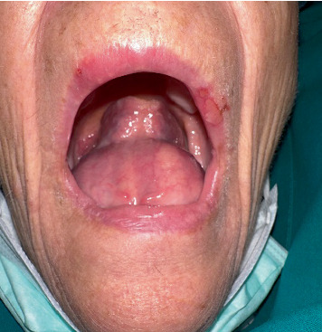
Aim: The aim of the study is to present a case of a dermoid cyst in the oral floor of an 80-year-old female patient and discuss its clinical presentation, diagnostic approach, and treatment. Methods: A retrospective case study was conducted at the Department of Maxillofacial Surgery, Aulss 2 Marca Trevigiana. The patient, with a medical history of hypertension, presented with a swelling in the oral floor. A clinical examination was followed by a facial CT scan with and without contrast medium to evaluate the cystic lesion. The patient underwent surgical excision under general anesthesia, with subsequent histopathological analysis. Results: The CT scan revealed a round, homogeneous, hyperdense formation with no enhancement after contrast administration. Surgical excision was performed using an intraoral approach, and histopathology confirmed the diagnosis of a dermoid cyst with an epithelial lining of predominantly squamous type. The patient experienced no complications, and follow-up at 9 days and 2 months showed complete recovery. Conclusion: A dermoid cyst of the oral cavity, though rare in adults, should be considered in the differential diagnosis of oral floor swellings. Early diagnosis through clinical examination and imaging, followed by surgical excision, provides an effective and curative treatment.
期刊介绍:
Case Reports in Dentistry is a peer-reviewed, Open Access journal that publishes case reports and case series in all areas of dentistry, including periodontal diseases, dental implants, oral pathology, as well as oral and maxillofacial surgery.

 求助内容:
求助内容: 应助结果提醒方式:


