Pan An, Qiang Tian, Shijun Duan, Xiulong Feng, Yu Han, Yaoning Wei, Wen Wang, Qian Yin
{"title":"The Imaging Evaluation of Left Atrial Appendage: CT Large-Spiral Arterial Late Scan.","authors":"Pan An, Qiang Tian, Shijun Duan, Xiulong Feng, Yu Han, Yaoning Wei, Wen Wang, Qian Yin","doi":"10.2147/IJGM.S537696","DOIUrl":null,"url":null,"abstract":"<p><strong>Purpose: </strong>Compared with retrospective ECG-gated arterial phase scan, to investigate the clinical application value of dual-source CT large-spiral arterial late scan in the imaging evaluation of left atrial appendage (LAA).</p><p><strong>Patients and methods: </strong>A total of 108 patients requiring LAA CT angiography (CTA) due to atrial fibrillation (AF) were selected from September 2024 to December 2024, including 52 patients in group A (Flash large-spiral arterial late scan) and 56 patients in group B (retrospective ECG-gated arterial phase scan). All patients underwent double-phase scan. The interval between the two periods is 30s. Clinical data of patients were collected, the scanning range and radiation dose received were recorded. The CT values of LAA, ascending aorta (AA) at the same level and left atrium at the largest level were measured. At the same time, evaluate other meaningful lesions within the scan range.</p><p><strong>Results: </strong>The difference between the two groups of scanning range and radiation dose was statistically significant (<i>P</i> < 0.05). There was statistical significance in the subjective evaluation and objective evaluation of LAA filling at the first phase scanning of the two groups (<i>P</i> < 0.05). The detection of other meaningful lesions in the scanning range of group A was significantly higher than that of group B.</p><p><strong>Conclusion: </strong>By adopting the three-generation dual-source FLASH large-pitch arterial late scan mode, the complete filling rate of the LAA was significantly improved. Not only shortened the examination time, reduced the radiation dose, but also increased the detection rate of other significant lesions within the scanning range for the patients.</p>","PeriodicalId":14131,"journal":{"name":"International Journal of General Medicine","volume":"18 ","pages":"4965-4973"},"PeriodicalIF":2.0000,"publicationDate":"2025-09-01","publicationTypes":"Journal Article","fieldsOfStudy":null,"isOpenAccess":false,"openAccessPdf":"https://www.ncbi.nlm.nih.gov/pmc/articles/PMC12412602/pdf/","citationCount":"0","resultStr":null,"platform":"Semanticscholar","paperid":null,"PeriodicalName":"International Journal of General Medicine","FirstCategoryId":"3","ListUrlMain":"https://doi.org/10.2147/IJGM.S537696","RegionNum":4,"RegionCategory":"医学","ArticlePicture":[],"TitleCN":null,"AbstractTextCN":null,"PMCID":null,"EPubDate":"2025/1/1 0:00:00","PubModel":"eCollection","JCR":"Q2","JCRName":"MEDICINE, GENERAL & INTERNAL","Score":null,"Total":0}
引用次数: 0
Abstract
Purpose: Compared with retrospective ECG-gated arterial phase scan, to investigate the clinical application value of dual-source CT large-spiral arterial late scan in the imaging evaluation of left atrial appendage (LAA).
Patients and methods: A total of 108 patients requiring LAA CT angiography (CTA) due to atrial fibrillation (AF) were selected from September 2024 to December 2024, including 52 patients in group A (Flash large-spiral arterial late scan) and 56 patients in group B (retrospective ECG-gated arterial phase scan). All patients underwent double-phase scan. The interval between the two periods is 30s. Clinical data of patients were collected, the scanning range and radiation dose received were recorded. The CT values of LAA, ascending aorta (AA) at the same level and left atrium at the largest level were measured. At the same time, evaluate other meaningful lesions within the scan range.
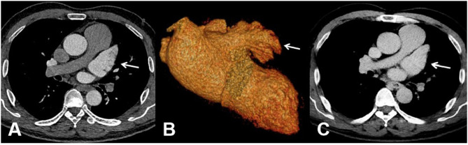
Results: The difference between the two groups of scanning range and radiation dose was statistically significant (P < 0.05). There was statistical significance in the subjective evaluation and objective evaluation of LAA filling at the first phase scanning of the two groups (P < 0.05). The detection of other meaningful lesions in the scanning range of group A was significantly higher than that of group B.
Conclusion: By adopting the three-generation dual-source FLASH large-pitch arterial late scan mode, the complete filling rate of the LAA was significantly improved. Not only shortened the examination time, reduced the radiation dose, but also increased the detection rate of other significant lesions within the scanning range for the patients.
期刊介绍:
The International Journal of General Medicine is an international, peer-reviewed, open access journal that focuses on general and internal medicine, pathogenesis, epidemiology, diagnosis, monitoring and treatment protocols. The journal is characterized by the rapid reporting of reviews, original research and clinical studies across all disease areas.
A key focus of the journal is the elucidation of disease processes and management protocols resulting in improved outcomes for the patient. Patient perspectives such as satisfaction, quality of life, health literacy and communication and their role in developing new healthcare programs and optimizing clinical outcomes are major areas of interest for the journal.
As of 1st April 2019, the International Journal of General Medicine will no longer consider meta-analyses for publication.

 求助内容:
求助内容: 应助结果提醒方式:


