Ali M Komai, Saliha Musovic, Kaj Stenlöf, Ludger Grote, Ding Zou, Jan Hedner
{"title":"Reduction of carbonic anhydrase activity is associated with amelioration of obstructive sleep apnea.","authors":"Ali M Komai, Saliha Musovic, Kaj Stenlöf, Ludger Grote, Ding Zou, Jan Hedner","doi":"10.1007/s11325-025-03430-z","DOIUrl":null,"url":null,"abstract":"<p><strong>Background: </strong>The carbonic anhydrase (CA) enzyme plays an important role in the equilibration of carbon dioxide and bicarbonate (HCO<sub>3</sub>) under the production of H<sup>+</sup> ions. Emerging evidence suggest that CA activity may play a fundamental regulatory role on respiratory control mechanisms in obstructive sleep apnea (OSA). Clinical trials suggest that CA inhibitors significantly reduce OSA.</p><p><strong>Methods: </strong>Data from three separate cohorts of healthy volunteers and patients with OSA were used to quantify CA activity in whole blood and cerebrospinal fluid (CSF). The influence of the CA inhibitory drugs acetazolamide and sulthiame, on CA activity in-vitro/in-vivo, was assessed. The association between CA-inhibitor plasma concentration and HCO<sub>3</sub>, as well as the influence of HCO<sub>3</sub> on the apnea-hypopnea severity was determined.</p><p><strong>Results: </strong>Stability of CA activity in stored blood was high. CA activity in whole blood contained five times higher activity compared with CSF. The CA-inhibitory drugs dose-dependently reduced CA activity in-vitro/in-vivo. The CA inhibitor sulthiame reduced venous HCO<sub>3</sub> concentration (P = 0.022). The reduction of HCO<sub>3</sub> was linked to improvement of OSA (P = 0.040).</p><p><strong>Conclusions: </strong>CA-inhibitory drugs reduced CA activity in whole blood suggesting a therapeutic role of CA inhibition in OSA. The findings also suggest that an activated CA system may constitute a pathophysiological mechanism in some forms of OSA.</p><p><strong>Clinical trial registration: </strong>N/A.</p>","PeriodicalId":520777,"journal":{"name":"Sleep & breathing = Schlaf & Atmung","volume":"29 5","pages":"278"},"PeriodicalIF":2.0000,"publicationDate":"2025-09-04","publicationTypes":"Journal Article","fieldsOfStudy":null,"isOpenAccess":false,"openAccessPdf":"https://www.ncbi.nlm.nih.gov/pmc/articles/PMC12411314/pdf/","citationCount":"0","resultStr":null,"platform":"Semanticscholar","paperid":null,"PeriodicalName":"Sleep & breathing = Schlaf & Atmung","FirstCategoryId":"1085","ListUrlMain":"https://doi.org/10.1007/s11325-025-03430-z","RegionNum":0,"RegionCategory":null,"ArticlePicture":[],"TitleCN":null,"AbstractTextCN":null,"PMCID":null,"EPubDate":"","PubModel":"","JCR":"","JCRName":"","Score":null,"Total":0}
引用次数: 0
Abstract
Background: The carbonic anhydrase (CA) enzyme plays an important role in the equilibration of carbon dioxide and bicarbonate (HCO3) under the production of H+ ions. Emerging evidence suggest that CA activity may play a fundamental regulatory role on respiratory control mechanisms in obstructive sleep apnea (OSA). Clinical trials suggest that CA inhibitors significantly reduce OSA.
Methods: Data from three separate cohorts of healthy volunteers and patients with OSA were used to quantify CA activity in whole blood and cerebrospinal fluid (CSF). The influence of the CA inhibitory drugs acetazolamide and sulthiame, on CA activity in-vitro/in-vivo, was assessed. The association between CA-inhibitor plasma concentration and HCO3, as well as the influence of HCO3 on the apnea-hypopnea severity was determined.
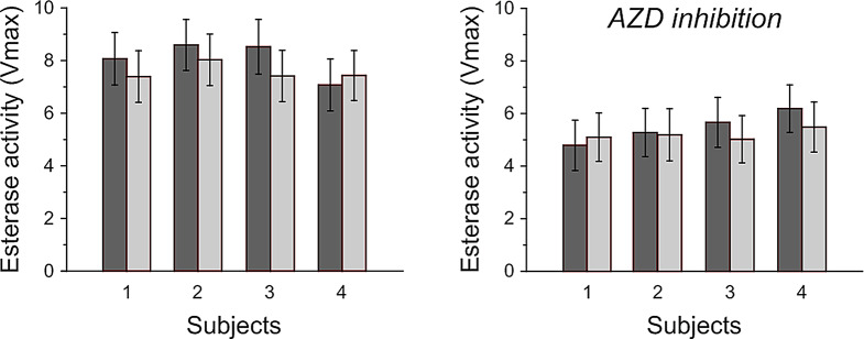
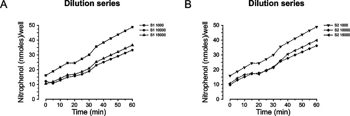
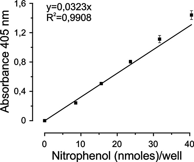
Results: Stability of CA activity in stored blood was high. CA activity in whole blood contained five times higher activity compared with CSF. The CA-inhibitory drugs dose-dependently reduced CA activity in-vitro/in-vivo. The CA inhibitor sulthiame reduced venous HCO3 concentration (P = 0.022). The reduction of HCO3 was linked to improvement of OSA (P = 0.040).
Conclusions: CA-inhibitory drugs reduced CA activity in whole blood suggesting a therapeutic role of CA inhibition in OSA. The findings also suggest that an activated CA system may constitute a pathophysiological mechanism in some forms of OSA.

 求助内容:
求助内容: 应助结果提醒方式:


