Bryce D Beutler, Daniel Antwi-Amoabeng, Dane Weinert, Ishan Shah, Mark B Ulanja, Alastair E Moody, Xiaomeng Lei, Alexander Lerner, Mark S Shiroishi, Reza Assadsangabi
{"title":"Prognostic value of arterial spin-labeling perfusion in anoxic brain injury: A retrospective cohort study.","authors":"Bryce D Beutler, Daniel Antwi-Amoabeng, Dane Weinert, Ishan Shah, Mark B Ulanja, Alastair E Moody, Xiaomeng Lei, Alexander Lerner, Mark S Shiroishi, Reza Assadsangabi","doi":"10.4329/wjr.v17.i8.111065","DOIUrl":null,"url":null,"abstract":"<p><strong>Background: </strong>Anoxic brain injury is a potentially lethal condition characterized by cerebral hypoperfusion and irreversible neuronal injury. Arterial spin-labeling (ASL) perfusion and diffusion-weighted imaging (DWI) magnetic resonance imaging (MRI) have been proposed as tools to detect cerebral ischemic changes and may aid in the assessment of anoxic injury.</p><p><strong>Aim: </strong>To explore the relationship between regional ASL perfusion patterns and clinical outcomes following cardiac arrest.</p><p><strong>Methods: </strong>We performed a retrospective review to identify patients with clinical suspicion of anoxic brain injury who underwent MRI within 15 days of cardiac arrest. Receiver operator characteristic (ROC) analysis and univariate logistic regression were used to evaluate associations between ASL perfusion scores, DWI signal intensity, and the following clinical features: (1) Myoclonus status epilepticus (MSE) within 24 hours; (2) Absent extensor or motor reflexes (EMR) at day 3 post-arrest; and (3) Absent brainstem reflexes (BSR) within 15 days.</p><p><strong>Results: </strong>Twenty-eight patients met inclusion criteria. Increased ASL signal in the left occipital lobe was significantly associated with MSE (<i>P</i> = 0.038), while a trend was observed between right frontal ASL signal and EMR (<i>P</i> = 0.078). ROC analysis showed that ASL scores ≥ 7 were associated with higher odds of absent BSR (OR 2.14, <i>P</i> = 0.53), though this did not reach statistical significance. DWI signal intensity did not show significant associations with clinical outcomes. The overall discriminatory performance of ASL for predicting outcomes was limited (AUC ≈ 0.52).</p><p><strong>Conclusion: </strong>This exploratory study suggests that regional ASL hyperperfusion, particularly in the left occipital and right frontal lobes, may be associated with adverse clinical signs following cardiac arrest. However, most findings did not reach statistical significance, and the study was underpowered to detect small-to-moderate effects. These preliminary results should be interpreted with caution and considered hypothesis-generating. Larger, prospective studies are warranted to clarify the prognostic value of ASL perfusion imaging in anoxic brain injury.</p>","PeriodicalId":23819,"journal":{"name":"World journal of radiology","volume":"17 8","pages":"111065"},"PeriodicalIF":1.5000,"publicationDate":"2025-08-28","publicationTypes":"Journal Article","fieldsOfStudy":null,"isOpenAccess":false,"openAccessPdf":"https://www.ncbi.nlm.nih.gov/pmc/articles/PMC12400257/pdf/","citationCount":"0","resultStr":null,"platform":"Semanticscholar","paperid":null,"PeriodicalName":"World journal of radiology","FirstCategoryId":"1085","ListUrlMain":"https://doi.org/10.4329/wjr.v17.i8.111065","RegionNum":0,"RegionCategory":null,"ArticlePicture":[],"TitleCN":null,"AbstractTextCN":null,"PMCID":null,"EPubDate":"","PubModel":"","JCR":"Q3","JCRName":"RADIOLOGY, NUCLEAR MEDICINE & MEDICAL IMAGING","Score":null,"Total":0}
引用次数: 0
Abstract
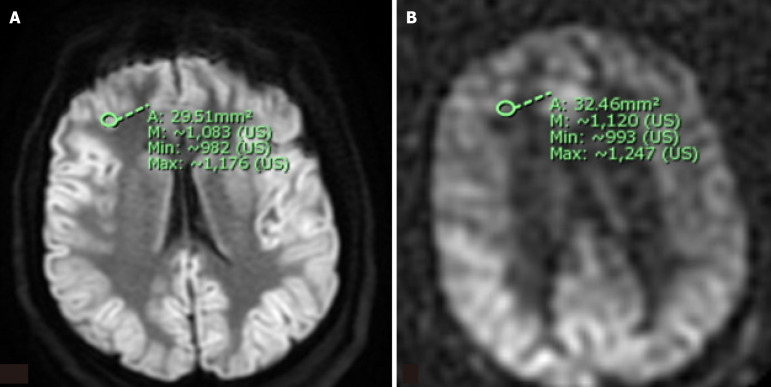
Background: Anoxic brain injury is a potentially lethal condition characterized by cerebral hypoperfusion and irreversible neuronal injury. Arterial spin-labeling (ASL) perfusion and diffusion-weighted imaging (DWI) magnetic resonance imaging (MRI) have been proposed as tools to detect cerebral ischemic changes and may aid in the assessment of anoxic injury.
Aim: To explore the relationship between regional ASL perfusion patterns and clinical outcomes following cardiac arrest.
Methods: We performed a retrospective review to identify patients with clinical suspicion of anoxic brain injury who underwent MRI within 15 days of cardiac arrest. Receiver operator characteristic (ROC) analysis and univariate logistic regression were used to evaluate associations between ASL perfusion scores, DWI signal intensity, and the following clinical features: (1) Myoclonus status epilepticus (MSE) within 24 hours; (2) Absent extensor or motor reflexes (EMR) at day 3 post-arrest; and (3) Absent brainstem reflexes (BSR) within 15 days.
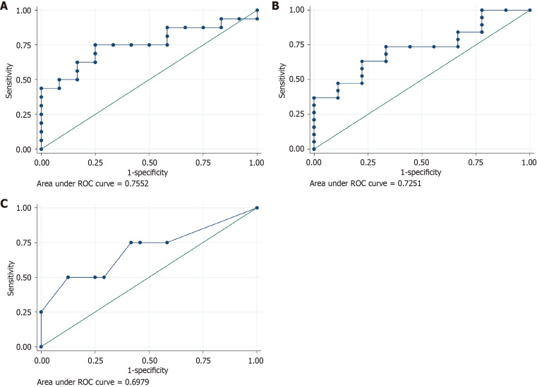
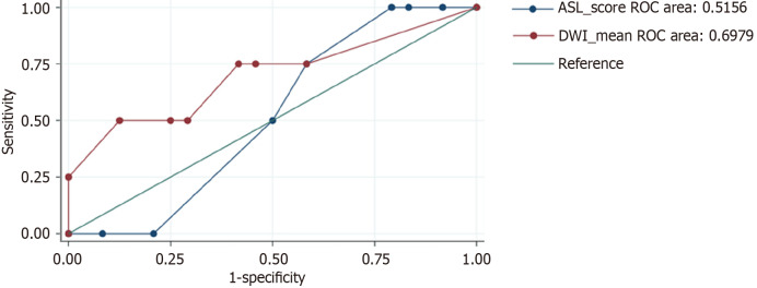
Results: Twenty-eight patients met inclusion criteria. Increased ASL signal in the left occipital lobe was significantly associated with MSE (P = 0.038), while a trend was observed between right frontal ASL signal and EMR (P = 0.078). ROC analysis showed that ASL scores ≥ 7 were associated with higher odds of absent BSR (OR 2.14, P = 0.53), though this did not reach statistical significance. DWI signal intensity did not show significant associations with clinical outcomes. The overall discriminatory performance of ASL for predicting outcomes was limited (AUC ≈ 0.52).
Conclusion: This exploratory study suggests that regional ASL hyperperfusion, particularly in the left occipital and right frontal lobes, may be associated with adverse clinical signs following cardiac arrest. However, most findings did not reach statistical significance, and the study was underpowered to detect small-to-moderate effects. These preliminary results should be interpreted with caution and considered hypothesis-generating. Larger, prospective studies are warranted to clarify the prognostic value of ASL perfusion imaging in anoxic brain injury.

 求助内容:
求助内容: 应助结果提醒方式:


