{"title":"Role of imaging in chronic otitis media and its complications.","authors":"Kemal Bugra Memis, Sonay Aydin","doi":"10.4329/wjr.v17.i8.109447","DOIUrl":null,"url":null,"abstract":"<p><p>Chronic otitis media (COM) is a long-standing inflammatory condition affecting the middle ear and mastoid cavity, often resulting in progressive structural damage and functional deficits. Radiological imaging is fundamental in diagnosing the disease, assessing its severity, and identifying possible complications. The literature indicates that the prevalence rates of extracranial and intracranial complications range from 0.69% to 5%, while the mortality rate for intracranial complications is 26%. While magnetic resonance imaging is particularly useful in distinguishing soft tissue abnormalities and detecting intracranial extensions like meningitis, brain abscess, and sigmoid sinus thrombosis, high-resolution computed tomography remains the preferred modality for evaluating bony erosion, cholesteatoma, and mastoid involvement. Key complications such as ossicular chain destruction, facial nerve damage, and labyrinthine fistulae can be precisely identified using advanced imaging modalities, allowing for timely and effective surgical intervention. This minireview underscores the essential role of radiology in both diagnosing and managing COM, highlighting critical imaging findings that facilitate early detection and inform treatment decisions. A collaborative approach among radiologists, otolaryngologists, and infectious disease specialists is crucial for improving clinical outcomes in affected patients.</p>","PeriodicalId":23819,"journal":{"name":"World journal of radiology","volume":"17 8","pages":"109447"},"PeriodicalIF":1.5000,"publicationDate":"2025-08-28","publicationTypes":"Journal Article","fieldsOfStudy":null,"isOpenAccess":false,"openAccessPdf":"https://www.ncbi.nlm.nih.gov/pmc/articles/PMC12400283/pdf/","citationCount":"0","resultStr":null,"platform":"Semanticscholar","paperid":null,"PeriodicalName":"World journal of radiology","FirstCategoryId":"1085","ListUrlMain":"https://doi.org/10.4329/wjr.v17.i8.109447","RegionNum":0,"RegionCategory":null,"ArticlePicture":[],"TitleCN":null,"AbstractTextCN":null,"PMCID":null,"EPubDate":"","PubModel":"","JCR":"Q3","JCRName":"RADIOLOGY, NUCLEAR MEDICINE & MEDICAL IMAGING","Score":null,"Total":0}
引用次数: 0
Abstract
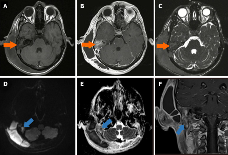
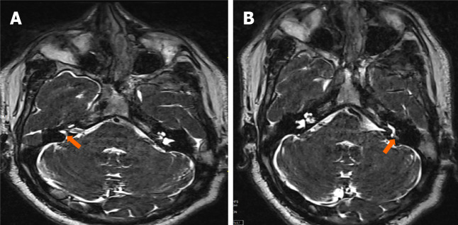
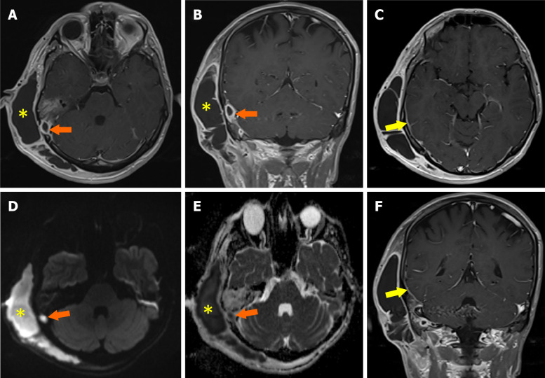
Chronic otitis media (COM) is a long-standing inflammatory condition affecting the middle ear and mastoid cavity, often resulting in progressive structural damage and functional deficits. Radiological imaging is fundamental in diagnosing the disease, assessing its severity, and identifying possible complications. The literature indicates that the prevalence rates of extracranial and intracranial complications range from 0.69% to 5%, while the mortality rate for intracranial complications is 26%. While magnetic resonance imaging is particularly useful in distinguishing soft tissue abnormalities and detecting intracranial extensions like meningitis, brain abscess, and sigmoid sinus thrombosis, high-resolution computed tomography remains the preferred modality for evaluating bony erosion, cholesteatoma, and mastoid involvement. Key complications such as ossicular chain destruction, facial nerve damage, and labyrinthine fistulae can be precisely identified using advanced imaging modalities, allowing for timely and effective surgical intervention. This minireview underscores the essential role of radiology in both diagnosing and managing COM, highlighting critical imaging findings that facilitate early detection and inform treatment decisions. A collaborative approach among radiologists, otolaryngologists, and infectious disease specialists is crucial for improving clinical outcomes in affected patients.

 求助内容:
求助内容: 应助结果提醒方式:


