Anna Russo, Luca Marinelli, Vittorio Patanè, Marina Alessandrella, Maria Cristina Pezzella, Teresa Troiani, Gabriella Brancaccio, Camila Scharf, Giuseppe Argenziano, Salvatore Cappabianca, Alfonso Reginelli
{"title":"Whole-body magnetic resonance imaging for cutaneous melanoma staging: A scientific review.","authors":"Anna Russo, Luca Marinelli, Vittorio Patanè, Marina Alessandrella, Maria Cristina Pezzella, Teresa Troiani, Gabriella Brancaccio, Camila Scharf, Giuseppe Argenziano, Salvatore Cappabianca, Alfonso Reginelli","doi":"10.5306/wjco.v16.i8.109206","DOIUrl":null,"url":null,"abstract":"<p><strong>Background: </strong>Cutaneous melanoma is an aggressive skin cancer with high metastatic potential. Accurate staging is critical to guide therapeutic strategies and improve prognosis. Whole-body magnetic resonance imaging (WB-MRI), particularly when combined with diffusion-weighted imaging (DWI), has emerged as promising tool for comprehensive, radiation-free assessment of metastatic spread.</p><p><strong>Aim: </strong>To systematically review the diagnostic performance and clinical utility of WB-MRI in the staging and restaging of cutaneous melanoma, with comparison to conventional imaging modalities such as computed tomography (CT) and positron emission tomography/CT (PET/CT).</p><p><strong>Methods: </strong>A systematic literature review was conducted using PubMed, Embase, Scopus and Web of Science databases for studies published in the last 10 years. Inclusion criteria focused on comparative diagnostic accuracy studies of WB-MRI <i>vs</i> CT and PET/CT for melanoma staging. The methodological quality of the studies was appraised using the QUADAS-2 tool.</p><p><strong>Results: </strong>Sixteen studies involving over 700 patients met the inclusion criteria. WB-MRI showed high sensitivity (73%-90%) and specificity (up to 98%) in detecting metastases, particularly in bone, liver and soft tissue. DWI enhanced lesion detection, and WB-MRI often influenced clinical management decisions. However, CT outperformed WB-MRI in identifying small pulmonary nodules. AI-assisted analysis and contrast-enhanced sequences further improved diagnostic confidence.</p><p><strong>Conclusion: </strong>WB-MRI represents a robust imaging modality for staging cutaneous melanoma, offering superior soft-tissue contrast and functional imaging without ionizing radiation. Its strengths lie in detecting bone, liver and brain metastases. Challenges include limited lung lesion detection, cost, and availability. Advances in artificial intelligence, Hybrid PET/MRY systems, and radiomics are poised to expand WB-MRI's role in personalized melanoma management.</p>","PeriodicalId":23802,"journal":{"name":"World journal of clinical oncology","volume":"16 8","pages":"109206"},"PeriodicalIF":3.2000,"publicationDate":"2025-08-24","publicationTypes":"Journal Article","fieldsOfStudy":null,"isOpenAccess":false,"openAccessPdf":"https://www.ncbi.nlm.nih.gov/pmc/articles/PMC12400218/pdf/","citationCount":"0","resultStr":null,"platform":"Semanticscholar","paperid":null,"PeriodicalName":"World journal of clinical oncology","FirstCategoryId":"1085","ListUrlMain":"https://doi.org/10.5306/wjco.v16.i8.109206","RegionNum":0,"RegionCategory":null,"ArticlePicture":[],"TitleCN":null,"AbstractTextCN":null,"PMCID":null,"EPubDate":"","PubModel":"","JCR":"Q3","JCRName":"ONCOLOGY","Score":null,"Total":0}
引用次数: 0
Abstract
Background: Cutaneous melanoma is an aggressive skin cancer with high metastatic potential. Accurate staging is critical to guide therapeutic strategies and improve prognosis. Whole-body magnetic resonance imaging (WB-MRI), particularly when combined with diffusion-weighted imaging (DWI), has emerged as promising tool for comprehensive, radiation-free assessment of metastatic spread.
Aim: To systematically review the diagnostic performance and clinical utility of WB-MRI in the staging and restaging of cutaneous melanoma, with comparison to conventional imaging modalities such as computed tomography (CT) and positron emission tomography/CT (PET/CT).
Methods: A systematic literature review was conducted using PubMed, Embase, Scopus and Web of Science databases for studies published in the last 10 years. Inclusion criteria focused on comparative diagnostic accuracy studies of WB-MRI vs CT and PET/CT for melanoma staging. The methodological quality of the studies was appraised using the QUADAS-2 tool.
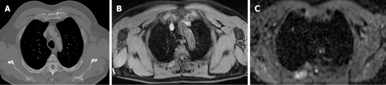
Results: Sixteen studies involving over 700 patients met the inclusion criteria. WB-MRI showed high sensitivity (73%-90%) and specificity (up to 98%) in detecting metastases, particularly in bone, liver and soft tissue. DWI enhanced lesion detection, and WB-MRI often influenced clinical management decisions. However, CT outperformed WB-MRI in identifying small pulmonary nodules. AI-assisted analysis and contrast-enhanced sequences further improved diagnostic confidence.
Conclusion: WB-MRI represents a robust imaging modality for staging cutaneous melanoma, offering superior soft-tissue contrast and functional imaging without ionizing radiation. Its strengths lie in detecting bone, liver and brain metastases. Challenges include limited lung lesion detection, cost, and availability. Advances in artificial intelligence, Hybrid PET/MRY systems, and radiomics are poised to expand WB-MRI's role in personalized melanoma management.
背景:皮肤黑色素瘤是一种具有高转移潜力的侵袭性皮肤癌。准确的分期对指导治疗策略和改善预后至关重要。全身磁共振成像(WB-MRI),特别是与扩散加权成像(DWI)相结合,已成为一种有前途的工具,用于全面,无辐射评估转移性扩散。目的:系统回顾WB-MRI在皮肤黑色素瘤分期和再分期中的诊断性能和临床应用,并与传统成像方式(如计算机断层扫描(CT)和正电子发射断层扫描/CT (PET/CT))进行比较。方法:采用PubMed、Embase、Scopus、Web of Science等数据库对近10年发表的研究进行系统文献综述。入选标准侧重于对比WB-MRI与CT和PET/CT对黑色素瘤分期的诊断准确性研究。使用QUADAS-2工具对研究的方法学质量进行评价。结果:16项涉及700多例患者的研究符合纳入标准。WB-MRI在检测转移方面显示出高灵敏度(73%-90%)和特异性(高达98%),特别是在骨、肝脏和软组织中。DWI增强病变检测,而WB-MRI通常影响临床管理决策。然而,CT在识别小肺结节方面优于WB-MRI。人工智能辅助分析和对比增强序列进一步提高了诊断的可信度。结论:WB-MRI为皮肤黑色素瘤分期提供了一种强大的成像方式,在没有电离辐射的情况下提供了优越的软组织对比和功能成像。它的优势在于检测骨、肝和脑转移。挑战包括有限的肺病变检测,成本和可用性。人工智能、PET/ mri混合系统和放射组学的进步,将扩大腰磁共振成像在个性化黑色素瘤管理中的作用。
期刊介绍:
The WJCO is a high-quality, peer reviewed, open-access journal. The primary task of WJCO is to rapidly publish high-quality original articles, reviews, editorials, and case reports in the field of oncology. In order to promote productive academic communication, the peer review process for the WJCO is transparent; to this end, all published manuscripts are accompanied by the anonymized reviewers’ comments as well as the authors’ responses. The primary aims of the WJCO are to improve diagnostic, therapeutic and preventive modalities and the skills of clinicians and to guide clinical practice in oncology. Scope: Art of Oncology, Biology of Neoplasia, Breast Cancer, Cancer Prevention and Control, Cancer-Related Complications, Diagnosis in Oncology, Gastrointestinal Cancer, Genetic Testing For Cancer, Gynecologic Cancer, Head and Neck Cancer, Hematologic Malignancy, Lung Cancer, Melanoma, Molecular Oncology, Neurooncology, Palliative and Supportive Care, Pediatric Oncology, Surgical Oncology, Translational Oncology, and Urologic Oncology.

 求助内容:
求助内容: 应助结果提醒方式:


