Shuai Ren, Bin Qin, Marcus J Daniels, Liang Zeng, Ying Tian, Zhong-Qiu Wang
{"title":"Developing and validating a computed tomography radiomics strategy to predict lymph node metastasis in pancreatic cancer.","authors":"Shuai Ren, Bin Qin, Marcus J Daniels, Liang Zeng, Ying Tian, Zhong-Qiu Wang","doi":"10.4329/wjr.v17.i8.109373","DOIUrl":null,"url":null,"abstract":"<p><strong>Background: </strong>Lymph node metastasis (LNM) is a key prognostic factor in pancreatic cancer (PC). Accurate preoperative prediction of LNM remains challenging. Radiomics offers a noninvasive method to extract quantitative imaging features that may aid in predicting LNM.</p><p><strong>Aim: </strong>To investigate the potential value of a computed tomography (CT)-based radiomics model in prediction of LNM in PC.</p><p><strong>Methods: </strong>A retrospective analysis was performed on 168 pathologically confirmed PC patients who underwent contrast-enhanced-CT. Among them, 107 cases had no LNM, while 61 cases had confirmed LNM. These patients were randomly divided into a training cohort (<i>n</i> = 135) and a validation cohort (<i>n</i> = 33). A total of 792 radiomics features were extracted, comprising 396 features from the arterial phase and another 396 from the portal venous phase. The Minimum Redundancy Maximum Relevance and Least Absolute Shrinkage and Selection Operator methods were used for feature selection and Radiomics model construction. The receiver operating characteristic curve was employed to assess the diagnostic potential of the model, and DeLong test was used to compare the area under the curve (AUC) values of the model.</p><p><strong>Results: </strong>Six radiomics features from the arterial phase and nine from the portal venous phase were selected. The Radscore model demonstrated strong predictive performance for LNM in both the training and test cohorts, with AUC values ranging from 0.86 to 0.94, sensitivity between 66.7% and 91.7%, specificity from 71.4% to 100.0%, accuracy between 78.8% and 91.1%, PPV ranging from 64.7% to 100.0%, and negative predictive value between 84.0% and 93.8%. No significant differences in AUC values were observed between the arterial and portal venous phases in either the training or test set.</p><p><strong>Conclusion: </strong>The preoperative CT-based radiomics model exhibited robust predictive capability for identifying LNM in PC.</p>","PeriodicalId":23819,"journal":{"name":"World journal of radiology","volume":"17 8","pages":"109373"},"PeriodicalIF":1.5000,"publicationDate":"2025-08-28","publicationTypes":"Journal Article","fieldsOfStudy":null,"isOpenAccess":false,"openAccessPdf":"https://www.ncbi.nlm.nih.gov/pmc/articles/PMC12400248/pdf/","citationCount":"0","resultStr":null,"platform":"Semanticscholar","paperid":null,"PeriodicalName":"World journal of radiology","FirstCategoryId":"1085","ListUrlMain":"https://doi.org/10.4329/wjr.v17.i8.109373","RegionNum":0,"RegionCategory":null,"ArticlePicture":[],"TitleCN":null,"AbstractTextCN":null,"PMCID":null,"EPubDate":"","PubModel":"","JCR":"Q3","JCRName":"RADIOLOGY, NUCLEAR MEDICINE & MEDICAL IMAGING","Score":null,"Total":0}
引用次数: 0
Abstract
Background: Lymph node metastasis (LNM) is a key prognostic factor in pancreatic cancer (PC). Accurate preoperative prediction of LNM remains challenging. Radiomics offers a noninvasive method to extract quantitative imaging features that may aid in predicting LNM.
Aim: To investigate the potential value of a computed tomography (CT)-based radiomics model in prediction of LNM in PC.
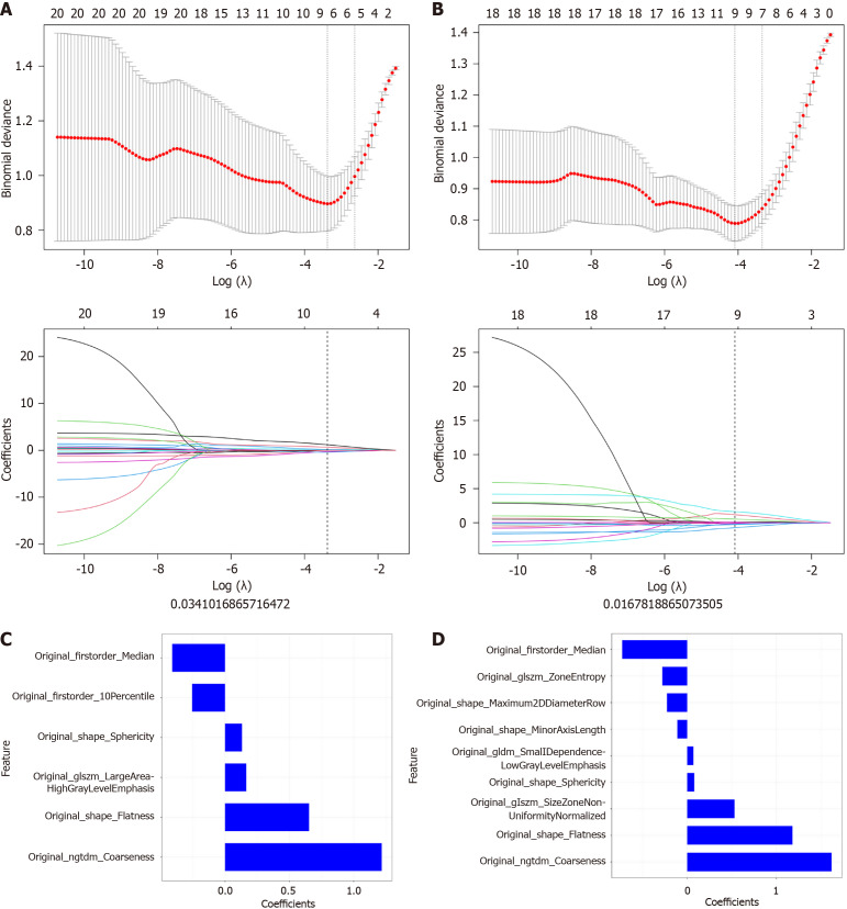
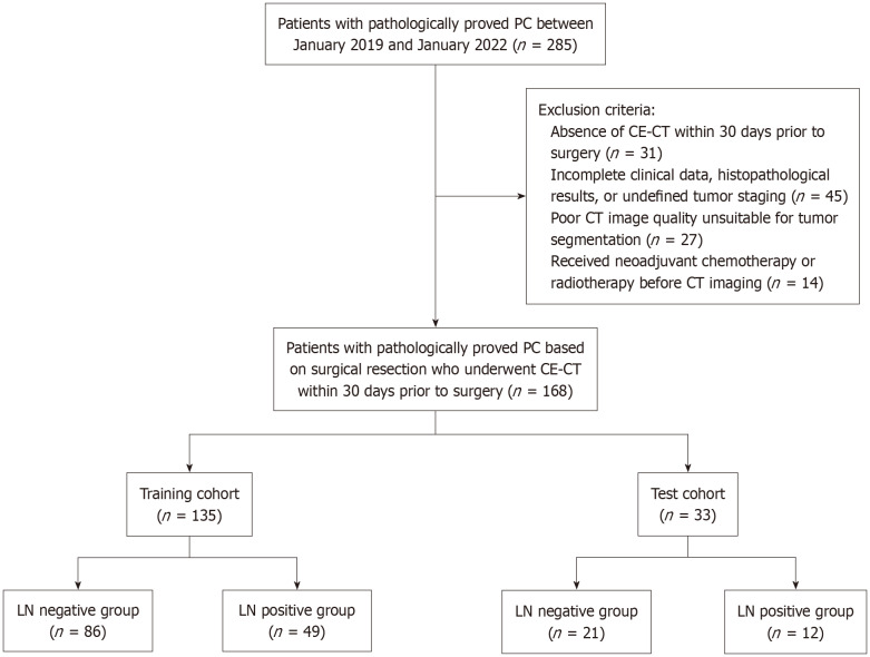
Methods: A retrospective analysis was performed on 168 pathologically confirmed PC patients who underwent contrast-enhanced-CT. Among them, 107 cases had no LNM, while 61 cases had confirmed LNM. These patients were randomly divided into a training cohort (n = 135) and a validation cohort (n = 33). A total of 792 radiomics features were extracted, comprising 396 features from the arterial phase and another 396 from the portal venous phase. The Minimum Redundancy Maximum Relevance and Least Absolute Shrinkage and Selection Operator methods were used for feature selection and Radiomics model construction. The receiver operating characteristic curve was employed to assess the diagnostic potential of the model, and DeLong test was used to compare the area under the curve (AUC) values of the model.
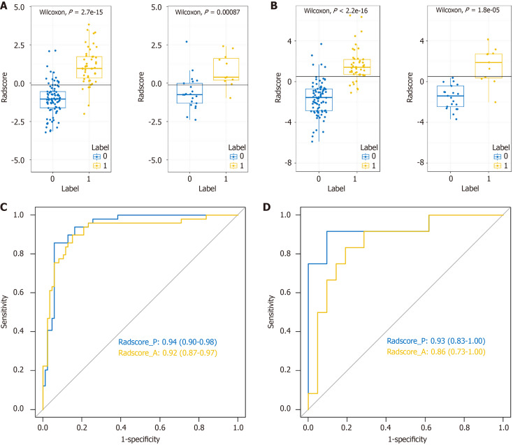
Results: Six radiomics features from the arterial phase and nine from the portal venous phase were selected. The Radscore model demonstrated strong predictive performance for LNM in both the training and test cohorts, with AUC values ranging from 0.86 to 0.94, sensitivity between 66.7% and 91.7%, specificity from 71.4% to 100.0%, accuracy between 78.8% and 91.1%, PPV ranging from 64.7% to 100.0%, and negative predictive value between 84.0% and 93.8%. No significant differences in AUC values were observed between the arterial and portal venous phases in either the training or test set.
Conclusion: The preoperative CT-based radiomics model exhibited robust predictive capability for identifying LNM in PC.

 求助内容:
求助内容: 应助结果提醒方式:


