Patrick Scherer, Nina Kurz-Mammri, Eva Graf, Thomas Papathemelis, Silke Haerteis, Cynthia Kohl
{"title":"Increase in Angiogenesis and Vascularization in Patient-Derived Endometriosis Tissue: Insights from a 3D In Vivo Model.","authors":"Patrick Scherer, Nina Kurz-Mammri, Eva Graf, Thomas Papathemelis, Silke Haerteis, Cynthia Kohl","doi":"10.1055/a-2649-6142","DOIUrl":null,"url":null,"abstract":"<p><strong>Aim: </strong>Endometriosis is a gynecological disorder characterized by endometrial-like tissue outside the uterus. This study evaluates the vascularization and proliferation of human endometriosis and endometrium tissues engrafted onto the chorioallantoic membrane of chicken embryos using immunohistochemistry and laser speckle contrast analysis imaging. For the assessment of clinical relevance, a comparison between laboratory and clinical data was performed.</p><p><strong>Material and methods: </strong>Tissue samples from 10 patients categorized by #Enzian scores and undergoing endometriosis surgery were investigated in the chorioallantoic membrane model. Hematoxylin-eosin staining and immunohistochemical markers, including CD10, cytokeratin, Ki67, and Caspase-3, assessed cellular structures, proliferation, and apoptosis. Changes in blood perfusion, implemented as a surrogate marker for angiogenesis and vascularization, were analyzed over three days using laser speckle contrast analysis. The fertilized chicken eggs used for the chorioallantoic membrane model were stratified for their gender utilizing an in ovo sexing technique.</p><p><strong>Results: </strong>Immunohistochemistry confirmed stromal and glandular cells in transplanted tissues. Ki67 indicated variable proliferation, while Caspase-3 identified apoptosis. Perfusion increased significantly in 75% of endometriosis samples. Endometrium from a patient with endometriosis showed increased perfusion, contrasting with stable perfusion in healthy endometrium. Higher #Enzian scores partly correlated with increased vascularization.</p><p><strong>Summary: </strong>The chorioallantoic membrane model is a viable platform for studying endometriosis vascularization and angiogenesis. Endometriosis tissue showed enhanced vascularization influenced by lesion size and anatomical location, offering insights into disease progression and therapeutic strategies.</p>","PeriodicalId":12481,"journal":{"name":"Geburtshilfe Und Frauenheilkunde","volume":"85 9","pages":"951-963"},"PeriodicalIF":1.9000,"publicationDate":"2025-07-22","publicationTypes":"Journal Article","fieldsOfStudy":null,"isOpenAccess":false,"openAccessPdf":"https://www.ncbi.nlm.nih.gov/pmc/articles/PMC12404789/pdf/","citationCount":"0","resultStr":null,"platform":"Semanticscholar","paperid":null,"PeriodicalName":"Geburtshilfe Und Frauenheilkunde","FirstCategoryId":"3","ListUrlMain":"https://doi.org/10.1055/a-2649-6142","RegionNum":4,"RegionCategory":"医学","ArticlePicture":[],"TitleCN":null,"AbstractTextCN":null,"PMCID":null,"EPubDate":"2025/9/1 0:00:00","PubModel":"eCollection","JCR":"Q2","JCRName":"OBSTETRICS & GYNECOLOGY","Score":null,"Total":0}
引用次数: 0
Abstract
Aim: Endometriosis is a gynecological disorder characterized by endometrial-like tissue outside the uterus. This study evaluates the vascularization and proliferation of human endometriosis and endometrium tissues engrafted onto the chorioallantoic membrane of chicken embryos using immunohistochemistry and laser speckle contrast analysis imaging. For the assessment of clinical relevance, a comparison between laboratory and clinical data was performed.
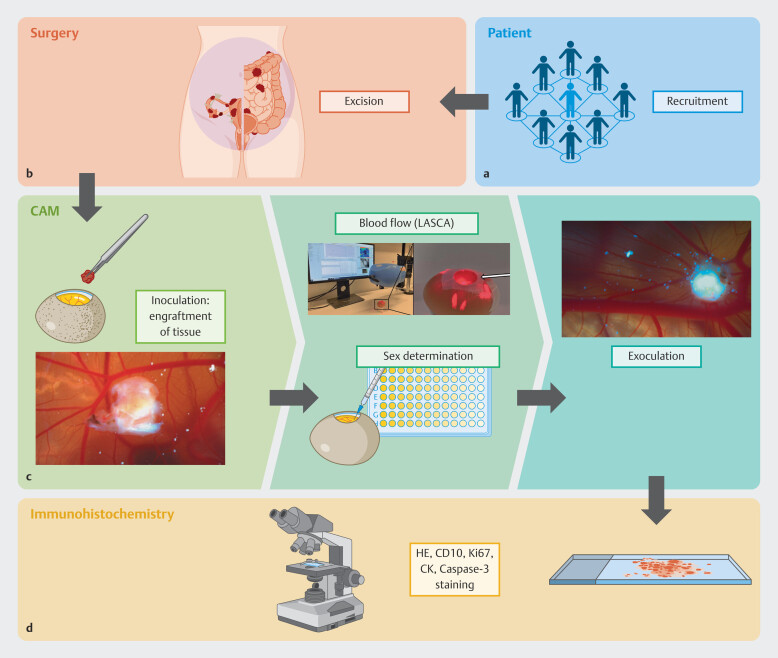
Material and methods: Tissue samples from 10 patients categorized by #Enzian scores and undergoing endometriosis surgery were investigated in the chorioallantoic membrane model. Hematoxylin-eosin staining and immunohistochemical markers, including CD10, cytokeratin, Ki67, and Caspase-3, assessed cellular structures, proliferation, and apoptosis. Changes in blood perfusion, implemented as a surrogate marker for angiogenesis and vascularization, were analyzed over three days using laser speckle contrast analysis. The fertilized chicken eggs used for the chorioallantoic membrane model were stratified for their gender utilizing an in ovo sexing technique.
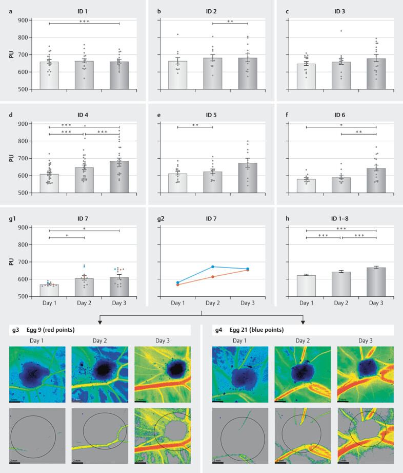
Results: Immunohistochemistry confirmed stromal and glandular cells in transplanted tissues. Ki67 indicated variable proliferation, while Caspase-3 identified apoptosis. Perfusion increased significantly in 75% of endometriosis samples. Endometrium from a patient with endometriosis showed increased perfusion, contrasting with stable perfusion in healthy endometrium. Higher #Enzian scores partly correlated with increased vascularization.
Summary: The chorioallantoic membrane model is a viable platform for studying endometriosis vascularization and angiogenesis. Endometriosis tissue showed enhanced vascularization influenced by lesion size and anatomical location, offering insights into disease progression and therapeutic strategies.
期刊介绍:
Geburtshilfe und Frauenheilkunde (GebFra) addresses the whole field of obstetrics and gynecology and is concerned with research as much as with clinical practice. In its scientific section, it publishes original articles, reviews and case reports in all fields of the discipline, namely
gynecological oncology, including oncology of the breast
obstetrics and perinatal medicine,
reproductive medicine,
and urogynecology.
GebFra invites the submission of original articles and review articles.
In addition, the journal publishes guidelines, statements and recommendations in cooperation with the DGGG, SGGG, OEGGG and the Arbeitsgemeinschaft der Wissenschaftlichen Medizinischen Fachgesellschaften (AWMF, Association of Scientific Medical Societies, www.awmf.org). Apart from the scientific section, Geburtshilfe und Frauenheilkunde has a news and views section that also includes discussions, book reviews and professional information.
Letters to the editors are welcome. If a letter discusses an article that has been published in our journal, the corresponding author of the article will be informed and invited to comment on the letter. The comment will be published along with the letter.

 求助内容:
求助内容: 应助结果提醒方式:


